
What is stress echocardiography and what is it used for?
Stress echocardiography allows the doctor to observe what changes occur in the heart when subjected to a stimulus, or ‘stress’, which can be either physical, i.e. induced by a patient’s muscular effort, or pharmacological
When the stress is pharmacological, the patient’s heart – which during the test is lying on the couch in the echocardiography laboratory – and its circulatory system are stimulated through the intravenous injection of specific drugs, which induce them to behave as if they were undergoing physical exertion.
Before performing pharmacological stress echo, the cardiologist always carries out an assessment of the heart by means of the normal echocardiogram, to consider whether the test is technically feasible and whether there are any contraindications to performing the test.
How is stress echocardiography performed?
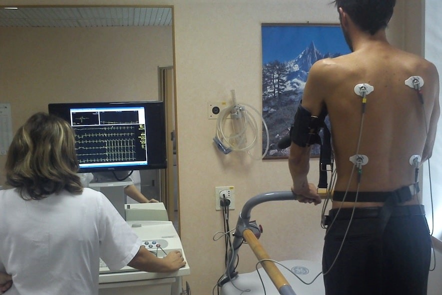
The patient is made to lie on a couch, on the left side, as for a normal transthoracic echocardiography.
Electrodes are applied to the chest to monitor the echocardiogram, and a sphygmomanometer, which monitors the blood pressure. At the same time, an intravenous drip (phleboclysis) administers, in a controlled dose, the drug that will cause the heart to undergo the same changes induced by physical exertion (Dobutamine) or changes in the coronary circulation (Dipyridamole or Atropine).
Other drugs deemed necessary by the cardiologist for the favourable outcome of the test (e.g. Atropine), or to reduce the heart rate at the end of the test (Metoprolol), may be administered intravenously via the drip.
The drip will then be removed at the end of the test.
Is Stress Echocardiography painful?
Stress echocardiography is not a painful test. In some cases, however, it may happen that patients with angina pectoris complain of the usual pain.
This is very important for the cardiologist, as he or she can check the correlation between the symptom and changes in cardiac function and the electrocardiogram.
When is the test interrupted?
The test is interrupted when the maximum dose of drug indicated for that particular patient has been injected; it may also be interrupted by the cardiologist, before completion of the injection protocol, in the event of the appearance of significant alterations in the electrocardiographic tracing or in the motility of the walls of the heart on the echocardiogram, for changes in the pressure parameters or when the patient complains of symptoms that the cardiologist considers significant.
Is Stress Echocardiography dangerous?
A dangerous arrhythmia, angina crisis, acute decompensation, myocardial infarction or cardiac arrest may occur during this type of test.
For this reason, in the laboratory where the test is performed, drugs and instruments are always available that can make each of these rare complications recede as quickly as possible.
The incidence of complications is however the same as for a normal exercise test on a treadmill or exercise bike, or a scintigraphy.
Test duration
30 minutes
Read Also
Emergency Live Even More…Live: Download The New Free App Of Your Newspaper For IOS And Android
Stress Electrocardiogram (ECG): An Overview Of The Test
Twenty-Four-Hour Ambulatory Blood Pressure Monitoring: What Does It Consist Of?
Holter Blood Pressure: Everything You Need To Know About This Test
What Is The Dynamic Electrocardiogram ECG According To Holter?
ECG: Waveform Analysis In The Electrocardiogram
What Is An ECG And When To Do An Electrocardiogram
ST-Elevation Myocardial Infarction: What Is A STEMI?
ECG First Principles From Handwritten Tutorial Video

ECG Criteria, 3 Simple Rules From Ken Grauer – ECG Recognize VT
Defibrillator: What It Is, How It Works, Price, Voltage, Manual And External
Cardiac Arrhythmias: Atrial Fibrillation
Full Dynamic Electrocardiogram According To Holter: What Is It?
Cardiac Rhythm Restoration Procedures: Electrical Cardioversion
Hypertension: Symptoms, Risk Factors And Prevention
Organ Complications Of Hypertension
How To Conduct Antihypertensive Treatment? An Overview Of Drugs
Blood Pressure: What It Is And How To Measure It
Aetiological Classification Of Hypertension
Classification Of Hypertension According To Organ Damage
Essential Hypertension: Pharmacological Associations In Antihypertensive Therapy
Treatment Of High Blood Pressure
Heart Failure: Causes, Symptoms And Treatment
The Thousand Faces Of Vascular Disease
Blood Pressure: When Is It High And When Is It Normal?
Metabolic Syndrome: Why Not To Underestimate It
Endocrine And Metabolic Emergencies In Emergency Medicine
Drug Therapy For The Treatment Of High Blood Pressure
Assess Your Risk Of Secondary Hypertension: What Conditions Or Diseases Cause High Blood Pressure?
Holter Monitor: How Does It Work And When Is It Needed?
What Is Patient Pressure Management? An Overview
Head Up Tilt Test, How The Test That Investigates The Causes Of Vagal Syncope Works
Cardiac Syncope: What It Is, How It Is Diagnosed And Who It Affects
Holter Blood Pressure: What Is The ABPM (Ambulatory Blood Pressure Monitoring) For?
Sinus Tachycardia: What It Is And How To Treat It
Emergency Room Access: Neurology Emergencies