
Congenital heart disease: tricuspid atresia
Tricuspid atresia is a congenital heart defect that is caused by the failure of the tricuspid valve to develop
Normally, oxygen-poor blood returns to the right atrium via the hollow veins, will pass into the right ventricle, which pumps it through the pulmonary arteries to the lungs where it will be oxygenated.
The oxygen-rich blood will then return from the lungs to the left atrium, pass into the left ventricle, and be pumped into the aorta to reach the whole body.
In tricuspid atresia, on the other hand, the valve not being formed will see the presence of fibrous tissue that closes it preventing the passage of oxygen-poor blood from the right atrium to the right ventricle
The right ventricle is often small (hypoplastic) due to reduced blood flow already in the embryonic stage.
In tricuspid atresias, which are not life-threatening, there will be a defect in the septum separating the two atria or the septum separating the ventricles, but usually both.
This will lead to the mixing of non-oxygenated blood with oxygenated blood.
The ductus arteriosus Botallo is an artery that, in the embryonic stage will allow the passage of blood from the pulmonary arteries to the aorta, will tend to close in the first few hours of life.
Should it remain open, it will see the passage of blood from the aorta to the pulmonary arteries and lungs where it will become oxygenated.
Thus, the baby born with tricuspid atresia will have blood that cannot flow through the heart and lungs to pick up oxygen as it normally would; the lung will not be able to supply the rest of the body with the oxygen it needs.
THE RADIO OF RESCUERS AROUND THE WORLD? IT’S RADIOEMS: VISIT ITS BOOTH AT EMERGENCY EXPO
Tricuspid atresia affects about 5 in 100,000 live births, affecting males and females equally
Symptoms of tricuspid atresia will include: exhaustion, shortness of breath, cyanosis, slow growth.
Symptoms that will occur as early as the second week after birth and are the most common.
Other symptoms will be: swelling of the lower limbs, abdominal bloating, weight gain from fluid retention, and irregular heartbeat.
Tricuspid atresia will occur during fetal heart development
Heredity or Down syndrome may also affect, increasing the risk of congenital heart defects.
Children with tricuspid atresia will also have ventricular septal defect; blood, in this case, will flow through the hole and into the right ventricle, which will pump it to the lungs.
Generally, the causes of congenital heart defects are unknown, but there are factors that influence their increased risk including: the mother contracting viral diseases during pregnancy, the presence of a congenital heart defect in either parent, certain drug therapies, and the presence of Down syndrome.
It is critical to intervene quickly in order to avoid fatal complications, including:
- lack of oxygen to tissues, a life-threatening condition; increased production of red blood cells, to compensate for the lack of oxygen caused by atresia, but which could lead to clot formation, heart attack and/or stroke.
- Thanks to ultrasound, it will be possible to diagnose atresia even before birth.
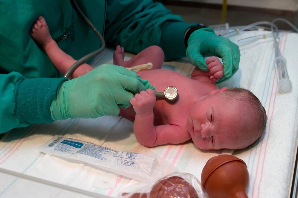
After birth, a heart defect will be thought of when the baby has bluish skin and breathing problems, or if a heart murmur is heard.
Through the echocardiogram, it will be possible to check for the absence of the tricuspid valve and a smaller-than-normal right ventricle; it will also be possible to measure how much blood flows between the right and left sides of the heart.
It will also be possible to check for heart defects such as atrial septal defect or ventricular septal defect.
Treatment of tricuspid atresia, will require surgery to ensure adequate blood flow through the heart and lungs so that the child’s body can receive the right amounts of oxygenated blood.
Multiple surgeries will obviously be used.
The surgical procedure that will be most frequently used will be the Fontan procedure, which will see the creation of a pathway for oxygen-depleted blood to flow properly into the pulmonary arteries.
It will be possible, however, to have the child undergo the Fontan procedure from the age of two.
Prior to Fontan’s surgery, drugs designed to help dilate blood vessels and keep the ductus arteriosus and foramen ovale open will be administered; antibiotics will be given before some dental procedures in order to prevent some bacteria from entering the bloodstream and reaching the heart and causing infective endocarditis.
Atrial septostomy will also be used to create, or enlarge, the opening between the cardiac atria in order to allow blood to flow from the right atrium to the left atrium.
With shunting instead, a bypass will be created starting at the aorta during the first 4 to 8 weeks of life.
With Glenn’s procedure, on the other hand, which will be performed around 6 months of age, a connection of the large veins that return blood to the heart to the pulmonary artery will be created; this procedure will reduce the workload of the left ventricle by decreasing the risk of damage.
For children who have undergone the Fontan procedure, the results are promising; however, complications may emerge over time that will require intervention with further procedures.
CHILD HEALTH: LEARN MORE ABOUT MEDICHILD BY VISITING THE BOOTH AT EMERGENCY EXPO
Read Also
Emergency Live Even More…Live: Download The New Free App Of Your Newspaper For IOS And Android
Bluish Color Of Baby’s Skin: Could Be Tricuspid Atresia
Supraventricular Tachycardia: Definition, Diagnosis, Treatment, And Prognosis
Neonatal/Pediatric Endotracheal Suctioning: General Characteristics Of The Procedure
Ventricular Aneurysm: How To Recognise It?
Atrial Fibrillation: Classification, Symptoms, Causes And Treatment

EMS: Pediatric SVT (Supraventricular Tachycardia) Vs Sinus Tachycardia
Atrioventricular (AV) Block: The Different Types And Patient Management
Pathologies Of The Left Ventricle: Dilated Cardiomyopathy
A Successful CPR Saves On A Patient With Refractory Ventricular Fibrillation
Atrial Fibrillation: Symptoms To Watch Out For
Atrial Fibrillation: Causes, Symptoms And Treatment
Difference Between Spontaneous, Electrical And Pharmacological Cardioversion
‘D’ For Deads, ‘C’ For Cardioversion! – Defibrillation And Fibrillation In Paediatric Patients
Inflammations Of The Heart: What Are The Causes Of Pericarditis?
Do You Have Episodes Of Sudden Tachycardia? You May Suffer From Wolff-Parkinson-White Syndrome (WPW)
Knowing Thrombosis To Intervene On The Blood Clot
Patient Procedures: What Is External Electrical Cardioversion?
Increasing The Workforce Of EMS, Training Laypeople In Using AED
Heart Attack: Characteristics, Causes And Treatment Of Myocardial Infarction
Altered Heart Rate: Palpitations
Heart: What Is A Heart Attack And How Do We Intervene?
Do You Have Heart Palpitations? Here Is What They Are And What They Indicate
Palpitations: What Causes Them And What To Do
Cardiac Arrest: What It Is, What The Symptoms Are And How To Intervene
Electrocardiogram (ECG): What It Is For, When It Is Needed
What Are The Risks Of WPW (Wolff-Parkinson-White) Syndrome
Heart Failure: Symptoms And Possible Treatments
What Is Heart Failure And How Can It Be Recognised?
Inflammations Of The Heart: Myocarditis, Infective Endocarditis And Pericarditis
Quickly Finding – And Treating – The Cause Of A Stroke May Prevent More: New Guidelines
Atrial Fibrillation: Symptoms To Watch Out For
Wolff-Parkinson-White Syndrome: What It Is And How To Treat It
Do You Have Episodes Of Sudden Tachycardia? You May Suffer From Wolff-Parkinson-White Syndrome (WPW)
What Is Takotsubo Cardiomyopathy (Broken Heart Syndrome)?
Heart Disease: What Is Cardiomyopathy?
Inflammations Of The Heart: Myocarditis, Infective Endocarditis And Pericarditis
Heart Murmurs: What It Is And When To Be Concerned
Broken Heart Syndrome Is On The Rise: We Know Takotsubo Cardiomyopathy
Heart Attack, Some Information For Citizens: What Is The Difference With Cardiac Arrest?
Heart Attack, Prediction And Prevention Thanks To Retinal Vessels And Artificial Intelligence
Full Dynamic Electrocardiogram According To Holter: What Is It?
In-Depth Analysis Of The Heart: Cardiac Magnetic Resonance Imaging (CARDIO – MRI)
Palpitations: What They Are, What Are The Symptoms And What Pathologies They Can Indicate
Cardiac Asthma: What It Is And What It Is A Symptom Of
Cardiac Rhythm Restoration Procedures: Electrical Cardioversion
Abnormal Electrical Activity Of The Heart: Ventricular Fibrillation
Gastro-Cardiac Syndrome (Or Roemheld Syndrome): Symptoms, Diagnosis And Treatment
Cardiac Arrhythmias: Atrial Fibrillation