
What is endoscopic transesophageal echocardiography?
Echocardiography is a method by which the heart and blood flow through the valves are studied using ultrasound
Unlike radiation used in radiology, ultrasound is harmless, so no precautions are necessary and the test can be performed on any patient countless times (even in pregnant women).
What is endoscopic transesophageal echocardiography used for?
Echocardiography allows us to obtain information about the contractility of the heart, the morphology of its valves, and the flow of blood in its cavities, both at rest and after exercise or taking a drug.
Transesophageal echocardiography represents a second-level test, generally indicated in cases where the transthoracic echocardiogram is deemed insufficient or uninterpretable with respect to the clinical question; in some cases it may be directly prescribed as the test of choice: presence of difficult-to-diagnose pathologies, such as rare congenital malformations, diseases of the thoracic aorta, or complex heart valve defects.
To perform the transesophageal echocardiogram, it is necessary to be fasting since midnight before the day of the test
Medications can be taken by trying to drink only the minimum amount sufficient to swallow the drugs.
If you have diabetes, it is important to consult with your doctor to determine the appropriate dose of insulin, which will obviously have to be reduced for fasting.
Who can perform endoscopic transesophageal echocardiography?
There are no particular contraindications to echocardiography: anyone can undergo the test.
How does endoscopic transesophageal echocardiography work?
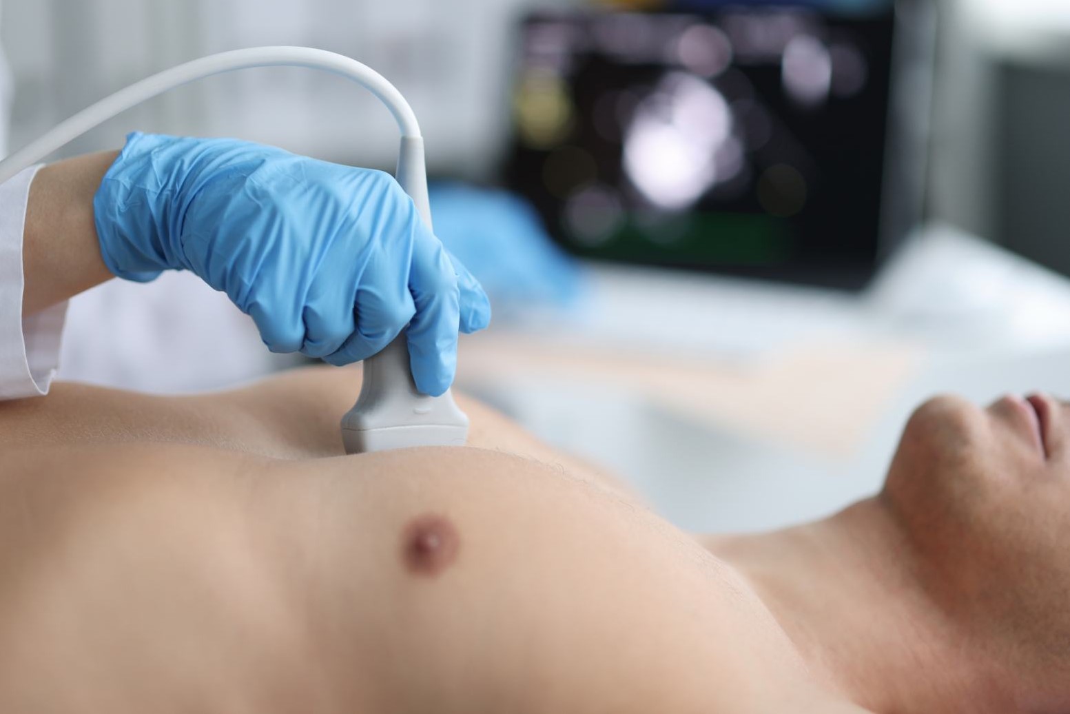
The patient must lie bare-chested on the sonographer’s bed, who will place electrodes on his chest.
Then the sonographer will spread a special gel on the patient’s chest and transducer, a probe that, when placed on the chest, emits ultrasound that, reflected and reprocessed by the equipment, allows the heart and its structures to be visualized.
The probe will be moved over the chest with gentle pressure.
The patient may be asked to remain still or breathe deeply.
At the end of the test, the electrodes will be removed and all that remains is to clean off the gel left on the chest.
The patient should remove any glasses and prostheses, lie on his side and position himself with torso and neck slightly flexed toward the legs.
He or she will then have to swallow a probe similar to that used for gastroscopy, inserted through a mouthpiece placed between the teeth.
The total duration of the test is about 10 to 15 minutes.
Is endoscopic transesophageal echocardiography painful or dangerous?
Transesophageal echocardiography is neither painful nor dangerous, but passing the probe through the mouth may generate some discomfort.
For this reason, to improve tolerance to the maneuver, the physician or nursing staff perform local anesthesia (spraying a spray down the throat), to which they may associate, in more sensitive patients, mild intravenous sedation.
In this case, since the state of alertness may be reduced, it will not be possible to drive or perform activities requiring special attention for at least 5-6 hours after the test.

Read Also
Emergency Live Even More…Live: Download The New Free App Of Your Newspaper For IOS And Android
Echo-Colour Doppler Of The Renal Arteries: What Does It Consist Of?
Echodoppler: What It Is And When To Perform It
Echo Doppler: What It Is And What It Is For
Doppler Test: When Is It Needed And How To Request It?
Fusion Prostate Biopsy: How The Examination Is Performed
What Is Needle Aspiration (Or Needle Biopsy Or Biopsy)?
What Is Echocolordoppler Of The Supra-Aortic Trunks (Carotids)?
What Is The Loop Recorder? Discovering Home Telemetry
Cardiac Holter, The Characteristics Of The 24-Hour Electrocardiogram
Peripheral Arteriopathy: Symptoms And Diagnosis
Endocavitary Electrophysiological Study: What Does This Examination Consist Of?
Cardiac Catheterisation, What Is This Examination?
Transesophageal Echocardiogram: What Does It Consist Of?
Venous Thrombosis: From Symptoms To New Drugs
Echotomography Of Carotid Axes
Echo- And CT-Guided Biopsy: What It Is And When It Is Needed
Echo-Doppler Of Vessels: Characteristics And Limitations Of The Method