
Echo-Doppler of vessels: characteristics and limitations of the method
Echo-Doppler of the vessels is a non-invasive diagnostic method, which is used to analyse arteries and veins. It is based on the same principles as the echocardiogram
What the Echo-Doppler of the vessels is used for
The Echo-Doppler of vessels is used to assess the morphology of blood vessels, their patency, the presence of malformations, narrowing (stenosis) or occlusions.
It can be used to explore both arteries and veins and now very often replaces other more invasive investigations such as arteriography and phlebography.
In addition, in the presence of vessel problems, the Echo-Doppler generally also allows us to define their severity and to set indications for treatment, which may be medical or surgical depending on the case.
How it is performed
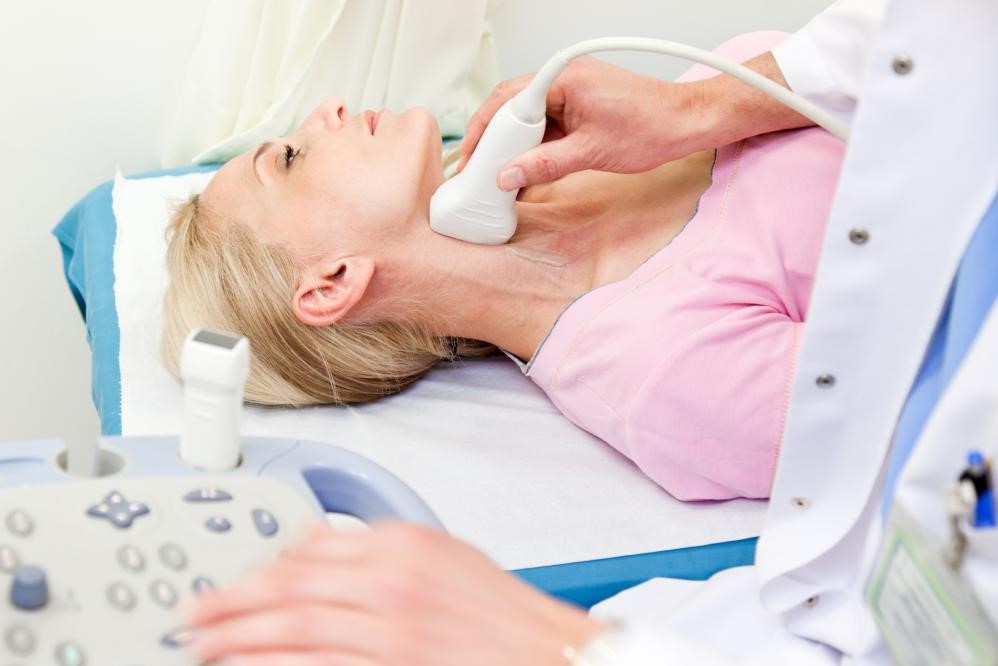
The test is generally conducted with the patient lying on a couch, although in selected cases it is more useful for the patient to remain standing.
It should be performed in a room with a comfortable temperature (22-25 C°) to prevent the cold from causing vasoconstriction (stiffening of the vessels), which decreases the volume of blood circulating in the superficial vessels, thus creating diagnostic problems.
The part of the body to be examined must be uncovered, taking care to remove especially tight clothing.
On the surface of the patient’s body, at the vessel to be analysed, the doctor moves a special probe capable of emitting ultrasound.
The ultrasound is reflected by the various structures of the body and, returning to the probe, creates an image of the vessel on the monitor.
Generally, a special gel is used which, when smeared on the patient’s skin surface, facilitates the passage of the ultrasound between the probe and the skin.
How long does the echo-doppler of the vessels last
The duration of the test varies from subject to subject, depending on the physical characteristics of the patient (longer in overweight or obese patients) and the difficulty of the clinical question.
On average, a test lasts about 15-20 minutes.
Due to the simplicity of its execution, the Vessel Echo-Doppler test can be performed in outpatients as well as in bedridden and critically ill patients.
Two major subgroups of pathologies that can be investigated with this method can be distinguished: those concerning arterial vessels and those concerning venous vessels.
Arterial vessel problems:
- Transcranial Echo-Doppler: this test is performed by placing the probe in various positions at the level of the patient’s head and is aimed at assessing blood flow in the cerebral vessels, particularly in the presence of a subarachnoid haemorrhage, a stroke or during neurosurgical interventions.
- Echo-Doppler of the neck vessels: this is very useful and widely used to diagnose problems in the carotid or vertebral artery, especially in subjects experiencing neurological symptoms such as dizziness, stroke or TIA. In these patients, narrowing (stenosis) or occlusion of the vessels in the neck may occur, requiring treatment (surgically or percutaneously) to prevent further aggravation of the condition. The Doppler test is also very important for following the progression of the disease in known patients and in the follow-up of operated patients.
- Aortic echo-Doppler: the aorta is the main arterial vessel, which starts from the heart and carries blood throughout the body, branching off into the other vessels. Quite frequently it develops age-related degenerative problems, such as diffuse calcifications. Sometimes it also dilates (aneurysm), thus running the risk of rupture, which is almost always fatal for the patient. By means of Echo-Doppler it is possible to assess the state of the aorta, measure its size and follow the increase in its diameter over time. When this increase reaches certain levels, the patient must undergo surgery to prevent rupture. Hence, this simple test is essential for diagnosing aneurysms and deciding when to undergo surgery. Given the importance of surgery, however, the patient often also undergoes a CT scan to confirm the diagnosis. The Doppler is then fundamental in following the patient after surgery.
- Echo-Doppler of the renal and mesenteric arteries: this test can be used to assess the flow in the arteries that carry blood to the kidneys and all the abdominal organs, although it is not always easy to perform due to the presence of fat. However, the method is facilitated if the patient is fasting. It is useful for identifying the presence of a renal artery stenosis, which can be a cause of high blood pressure, and for assessing the perfusion of other abdominal organs.
- Echo-Doppler of the lower limbs: in patients who complain of leg pain at rest or when walking, it is essential to rule out the presence of a stenosis of the arterial vessels of the lower limbs. Indeed, in the presence of a narrowing of these vessels, the amount of blood reaching the leg muscles is reduced. Under resting conditions, the blood demand from the muscles is low and therefore the blood flow through the narrowed (stenotic) vessels may be sufficient and the patient asymptomatic. Under stress, however, the muscle’s oxygen demands increase and the blood flow is thus inadequate. As a result, the muscle begins to suffer and the patient experiences severe pain, forcing him to stop exerting himself. To improve the situation, in the most serious cases, surgery (bypass) or angioplasty of the affected vessel must be performed. In all cases, an Echo-Doppler is used for diagnosis, which is able to define the extent of the narrowing, its progression, the need for surgery and to monitor the course after surgery.
Vein problems:
With regard to problems of the veins, the use of Eco-Doppler has its place in the diagnosis of venous insufficiency, which is one of the main causes of varicose veins (dilatations) of the lower limbs and phlebitis (inflammation of the veins), and in the diagnosis of venous thrombosis (occlusions of the veins) and thrombophlebitis (occlusions of the veins associated with inflammation).
Venous disorders cause local discomfort, as pain may develop, associated with swelling, heat, redness or ulcers of the affected limb, but more fearsome are the complications.
In the presence of venous thrombosis, the thrombotic material (blood clot that forms inside the vein) can suddenly detach from the site where it has formed and be transported in the bloodstream to the lungs, thus giving rise to pulmonary embolism, which can even be fatal.
This is why it is so important to make an early diagnosis and start the appropriate therapy as soon as possible, which can be medical (anticoagulant therapy, compression with elastic stockings) or, in more complex and risky cases, surgical.
Nowadays, almost all diagnostics of venous pathologies are performed by means of Echo-Doppler and phlebography (angiography of the affected venous district) is almost never necessary.
Advantages of the Echo-Doppler of the vessels
Compared to other methods, the Echo-Doppler of the vessels is relatively simple, absolutely non-invasive, quickly performed, inexpensive and can be performed at the patient’s bedside, thus being very useful even in critically ill patients who cannot be moved to perform more complex investigations.
It is painless and therefore well tolerated by everyone and the results are immediately interpretable.

Moreover, it is completely risk- and complication-free and can therefore be repeated frequently, making it very useful for assessing the evolution of a particular pathology or for serious checks after surgical or percutaneous interventions on vessels.
Side effects and risks
They are practically non-existent.
Limitations of the method
Not all vessels are well visualised with this method.
Moreover, in some subjects, particularly if they are obese, overweight or have significant skin oedema (accumulation of fluid), the difficulties increase.
For this reason, it is sometimes necessary to perform angiography of the affected vessels, especially in the presence of an indication for surgery.
Moreover, as with all ultrasound methods, the result depends very much on the skill and experience of the doctor performing the test.
Read Also
Emergency Live Even More…Live: Download The New Free App Of Your Newspaper For IOS And Android
Echodoppler: What It Is And When To Perform It
Echo Doppler: What It Is And What It Is For
Fusion Prostate Biopsy: How The Examination Is Performed
What Is Needle Aspiration (Or Needle Biopsy Or Biopsy)?
What Is Echocolordoppler Of The Supra-Aortic Trunks (Carotids)?
What Is The Loop Recorder? Discovering Home Telemetry
Cardiac Holter, The Characteristics Of The 24-Hour Electrocardiogram
Peripheral Arteriopathy: Symptoms And Diagnosis
Endocavitary Electrophysiological Study: What Does This Examination Consist Of?
Cardiac Catheterisation, What Is This Examination?
Transesophageal Echocardiogram: What Does It Consist Of?
Venous Thrombosis: From Symptoms To New Drugs
Echotomography Of Carotid Axes
Echo- And CT-Guided Biopsy: What It Is And When It Is Needed