
Congenital cyanogenic heart disease: tetralogy of Fallot
Tetralogy of Fallot is a congenital cyanogenic heart disease characterised by interventricular septal defect associated with obstruction of blood flow to the lungs
Tetralogy of Fallot is the most frequent cyanogenic congenital heart disease
Cyanosis is due to a reduction in circulating oxygen in the blood.
It mainly consists of these anatomical features:
- Communication between the two ventricles, the two pumping parts of the heart, thus defect of the ventricular septum;
- the biventricular origin of the aorta, which straddles the two ventricles, above the interventricular defect;
- subvalvular and pulmonary valve narrowing, muscle enlargement of the right ventricle, as a consequence of the other defects.
In Tetralogy of Fallot, there is a large interventricular septal defect associated with pulmonary stenosis of varying degrees, thus obstructing blood flow to the lungs.
Less blood will be oxygenated, desaturated blood will take the pathway to the aorta through the communication between the ventricles.
In young patients, the severity of tetralogy of Fallot is related to the severity of the pulmonary stenosis, which affects the extent of cyanosis
If oxygen is low, or if children have hypercyanotic crises, surgical treatment is carried out in the first few months of the child’s life; if the child has few symptoms, surgical treatment is sometimes carried out later in childhood.
In most cases, Tetralogy of Fallot is not associated with genetic abnormalities, but in 30% of patients there may be syndromes such as Down syndrome or Di George syndrome.
The main symptom is cyanosis; the level of cyanosis is usually stable but may differ from one patient to another.
After the neonatal period, there may be crises due to low oxygen levels in the blood, called asphyctic crises, which are a consequence of the sudden increase in the obstruction of blood flow to the lungs.
Due to the reduction in the level of oxygen in the blood, the baby’s lips and skin will appear more cyanotic; the baby will at first become irritable and if severe cyanosis persists there may be loss of consciousness.
Asphytic crisis is an episode that can be dangerous and requires immediate medical attention.
Infants with tetralogy of Fallot usually have a heart murmur
A murmur that is a sound created by blood flow in narrowed leaking heart valves or abnormal heart structures.
Some infants may have life-threatening fits of hypercyanosis; the cyanosis will suddenly worsen following crying or evacuation.
The baby will have accelerated breathing and in some cases there will be loss of consciousness. During these episodes the heart murmur will disappear.
In most cases, in prenatal diagnosis, with a morphological ultrasound the gynaecologist will be able to raise a suspicion of Tetralogy of Fallot; this will be confirmed with a fetal echocardiogram, and in the event of diagnostic confirmation, amniocentesis will be performed to rule out the presence of genetic syndromes.
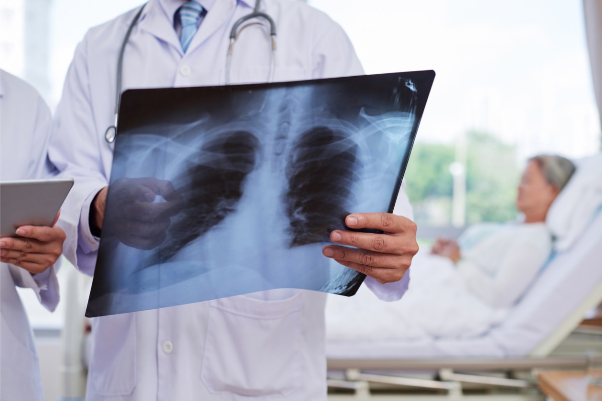
In the case of postnatal diagnosis, cyanosis and heart murmur may lead to suspicion of congenital heart disease, the echocardiogram being a rapid and non-invasive method is the most recommended diagnostic test.
In the treatment of Tetralogy of Fallot in the first few months of life, cardiac correction will be performed; before proceeding to definitive correction, a palliative pulmonary flow augmentation operation must be performed in infancy; the operative risk is extremely low.
If the child has hypercyanotic crisis, they will be able to breathe more easily if they bring their knees close to their chest; older children will also assume the same squatting position because it will help them push more blood to their lungs, making them feel better.
It may be helpful to administer oxygen.
If these measures do not work, morphine, intravenous beta blockers may be administered to improve blood flow to the lungs.
If children have low birth weight or complex defects, doctors may use less invasive procedures to maintain blood flow to the lungs while waiting for corrective surgery; for example, they may use a synthetic blood vessel (a shunt) to connect the aorta to an artery in the lungs.
This procedure directs the blood to the lungs so that it becomes oxygenated before being transported to the rest of the body.
Another option is cardiac catheterisation, in which a catheter with an expandable flexible tube (stent) at its tip is passed through a blood vessel in the leg to the heart.
The stent is expanded in the heart to widen the outflow to the lungs, increasing oxygen levels in the blood.
During corrective surgery, the defect in the interventricular septum is closed, the outflow pathway from the right ventricle and the stenotic pulmonary valve are dilated, and the pervious ductus arteriosus is closed.
Read Also
Emergency Live Even More…Live: Download The New Free App Of Your Newspaper For IOS And Android
Heart Abnormalities: What Is Tetralogy Of Fallot?
Tetralogy Of Fallot: Diagnosis, Prenatal Diagnosis And Differential Diagnosis
Tetralogy Of Fallot: Causes, Symptoms, Diagnosis, Treatment, Complications And Risks

Semeiotics Of The Heart: History In The Complete Cardiac Physical Examination
Electrical Cardioversion: What It Is, When It Saves A Life
Heart Murmur: What Is It And What Are The Symptoms?
Performing The Cardiovascular Objective Examination: The Guide
Branch Block: The Causes And Consequences To Take Into Account
Cardiopulmonary Resuscitation Manoeuvres: Management Of The LUCAS Chest Compressor
Supraventricular Tachycardia: Definition, Diagnosis, Treatment, And Prognosis
Identifying Tachycardias: What It Is, What It Causes And How To Intervene On A Tachycardia
Myocardial Infarction: Causes, Symptoms, Diagnosis And Treatment
Aortic Insufficiency: Causes, Symptoms, Diagnosis And Treatment Of Aortic Regurgitation
Congenital Heart Disease: What Is Aortic Bicuspidia?
Atrial Fibrillation: Definition, Causes, Symptoms, Diagnosis And Treatment
Ventricular Fibrillation Is One Of The Most Serious Cardiac Arrhythmias: Let’s Find Out About It
Atrial Flutter: Definition, Causes, Symptoms, Diagnosis And Treatment
What Is Echocolordoppler Of The Supra-Aortic Trunks (Carotids)?
What Is The Loop Recorder? Discovering Home Telemetry
Cardiac Holter, The Characteristics Of The 24-Hour Electrocardiogram
Peripheral Arteriopathy: Symptoms And Diagnosis
Endocavitary Electrophysiological Study: What Does This Examination Consist Of?
Cardiac Catheterisation, What Is This Examination?
Echo Doppler: What It Is And What It Is For
Transesophageal Echocardiogram: What Does It Consist Of?
Paediatric Echocardiogram: Definition And Use
Heart Diseases And Alarm Bells: Angina Pectoris
Fakes That Are Close To Our Hearts: Heart Disease And False Myths
Sleep Apnoea And Cardiovascular Disease: Correlation Between Sleep And Heart
Myocardiopathy: What Is It And How To Treat It?
Venous Thrombosis: From Symptoms To New Drugs
Cyanogenic Congenital Heart Disease: Transposition Of The Great Arteries
Heart Rate: What Is Bradycardia?
Consequences Of Chest Trauma: Focus On Cardiac Contusion