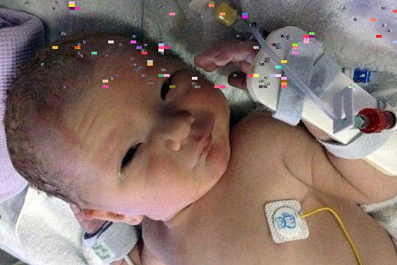
Tetralogy of Fallot: causes, symptoms, diagnosis, treatment, complications and risks
Tetralogy of Fallot, also known as “tetralogy of Fallot” or “blue baby syndrome” or “quadrilogy of Fallot” (hence the acronym TOF or ToF) is a congenital heart malformation, i.e. already present at birth, which classically has four anatomical elements that differentiate the heart of a newborn with tetralogy of Fallot from the heart of a healthy infant
If tetralogy of Fallot is also associated with a “patent foramen ovale” or a defect in the interatrial septum, the syndrome is called “pentalogy of Fallot”.
Spreading of tetralogy of Fallot
Tetralogy of Fallot is the most common cyanogenic congenital heart lesion in adults and accounts for 10% of all congenital heart diseases.
It may present to the physician before or, more commonly, after corrective or palliative surgery.
ECG EQUIPMENT? VISIT THE ZOLL BOOTH AT EMERGENCY EXPO
Causes and pathophysiology of blue baby syndrome
Tetralogy is the result of a malalignment of the aorto-pulmonary septum that divides the trunk of the arteries into the aorta and pulmonary artery during development, resulting in an anterior deviation of the aorta towards the pulmonary artery.
The four components of tetralogy of Fallot are as follows:
- aorta caving in on the interventricular septum;
- right ventricular outflow obstruction, which may occur at the valvular, subvalvular, supravalvular level or a combination of all three;
- membranous DIV (interventricular defect);
- hypertrophy of the right ventricle.
The DIV is usually large and allows the right and left ventricles to communicate freely.
The presence of right ventricular outflow obstruction is protective, preventing pressure and volume overload of the pulmonary circulation, which would result in fixed pulmonary hypertension.
The degree of right-left shunt depends on the degree of right ventricular outflow obstruction.
If pulmonary stenosis is mild, the left-right shunt is minimal and the patient remains achyanotic (pink tetralogy).
More often, pulmonary stenosis is severe and a large volume of poorly oxygenated blood is diverted into the systemic circulation resulting in cyanosis.
The cyanosis worsens with exercise, as the fall in systemic vascular resistance increases the degree of right-left shunt.
Tetralogy may also be associated with DIA (inter-atrial defect), muscular DIV, right aortic arch and other coronary anomalies.
A chromosomal deletion (22qll) is observed in 15% of cases, particularly in those with associated anomalies.
This deletion implies an increased risk of transmission of congenital heart disease to the offspring.
CHILD CARE PROFESSIONALS IN NETWOK: VISIT THE MEDICHILD BOOTH AT EMERGENCY EXPO
Diagnosis and symptoms of tetralogy of Fallot
If the mother is diabetic, a smoker, over 40 years old and the baby is premature with cyanosis and breathing difficulties, the doctor should consider tetralogy of Fallot as a likely possibility.
On physical examination there is cyanotic skin, digital hippocratism, prominent ejection murmur at the left sternal margin, reduced or absent P2.
The diagnostic doubt is then confirmed by:
- chest X-ray,
- electrocardiogram,
- echocardiogram and cardiac echocolordoppler.
The electrocardiogram shows hypertrophy of the right ventricle and abnormalities of the right atrium.
Chest X-ray shows characteristic shoehorn heart, small pulmonary artery, normal pulmonary vasculature.
Echocardiography determines the final diagnosis and usually provides sufficient information for planning surgical treatment.
In about half of all cases, tetralogy of Fallot is diagnosed before birth (prenatal diagnosis) by echocardiography.
Therapy
Surgical correction of tetralogy is usually performed in the neonatal period or in infancy and involves improvement of the right ventricular obstruction and patch closure of the DIV (interventricular defect).
After repair surgery, patients are at risk of residual stenosis or pulmonary insufficiency, which may lead to right ventricular dilatation and dysfunction and tricuspid insufficiency.
Aortic insufficiency is common after repair and may become clinically significant.
Residual AVD, right ventricular outflow tract aneurysms and sustained arrhythmias are also recognised complications.
Arrhythmias can be supraventricular or ventricular, can lead to haemodynamic compromise and contribute to an increased risk of sudden death.
Prolongation of QRS duration (up to >180 ms) on surface electrocardiogram (ECG) is a marker of increased risk of ventricular tachycardia and sudden death.
Palliative surgery may be performed in infancy to improve pulmonary blood flow.
Sometimes patients may choose not to undergo complete repair.
This palliation consists of creating a shunt between the systemic and pulmonary circulation, e.g. between the subclavian artery and the ipsilateral pulmonary artery (Blalock-Taussig shunt), which leads to an increase in pulmonary blood flow and an improvement in systemic blood oxygenation.
Various palliative shunts have been used for this purpose.
Although such procedures often result in long-term palliation of hypoxia, several complications can occur.
Shunts may become small as patients grow, or they may close spontaneously and lead to progressive cyanosis.
If the shunt is too large, the increased blood volume in the pulmonary circulation and left heart may lead to pulmonary congestion and progress to irreversible pulmonary vascular obstruction.
In patients who survive to adulthood, corrective surgery should still be attempted, but the surgical risk is greater due to the presence of right ventricular dysfunction.
All patients with tetralogy, even if the condition has been surgically corrected, should receive prophylaxis for endocarditis.
Read Also:
Emergency Live Even More…Live: Download The New Free App Of Your Newspaper For IOS And Android
Carotid Angioplasty And Stenting: What Are We Talking About?
Coronary Angioplasty, What To Do Post-Operatively?
Heart Patients And Heat: Cardiologist’s Advice For A Safe Summer
US EMS Rescuers To Be Assisted By Paediatricians Through Virtual Reality (VR)
Coronary Angioplasty, How Is The Procedure Performed?
Antiplatelet Drugs: Overview Of Their Usefulness


