
Diagnostic tests: what retinography and retinal fluorangiography are and when they are performed
Retinography and retinal fluorangiography are two particularly important tests for detecting vascular causes of eye diseases
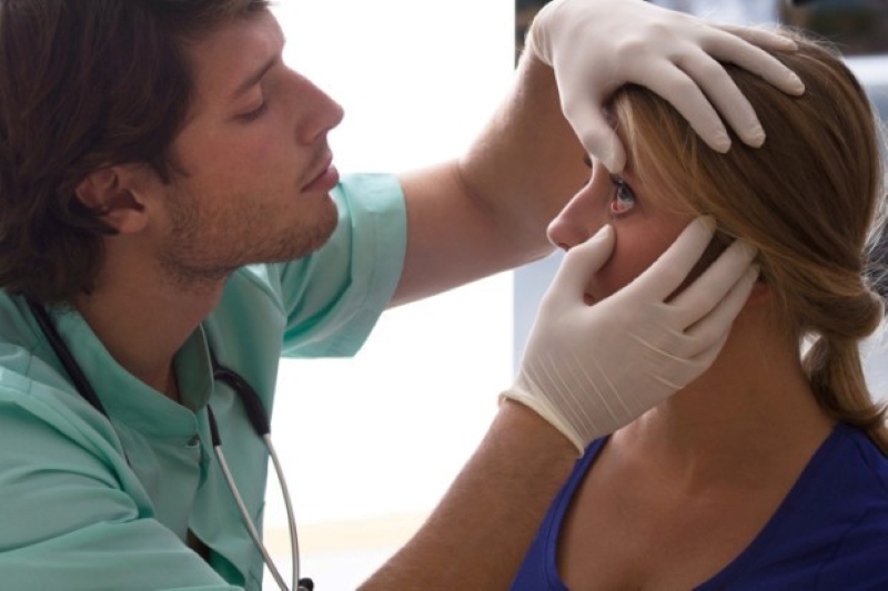
An eye examination is an essential moment in the detection of such diseases, and can save the patient from seriously disabling consequences.
What is retinography?
Retinography is a digitised photograph of the back of the eye.
This technique records the appearance of the patient’s retina, checking the results, detecting changes, studying them and recording them.
Specialist photography of the back of the eye consists of an adapted microscope connected to a flash camera that provides a magnified vertical view.
A classic camera provides a 30° and 50° view of the retinal area -with a 2.5x zoom-, with the central retina, macula and optic disc being the main structures that can be analysed.
There is also another form of retinography called wide field, which captures more than 200°.
This means that 80 per cent of the retina in a panoramic image that, in addition to being able to visualise the aforementioned structures, captures a complete image of the peripheral retina.
How is retinography performed?
Central retinography requires prior dilation of the pupil with eye drops that take effect within about 15 minutes and lead to blurred vision and glare for several hours.
Wide-field retinography does not require the use of mydriatic eye drops.
Both technologies provide a very fast test that is performed in the outpatient clinic in 10 minutes in the case of the module and less than 5 in the case of the latter, as only one photograph is required, which is taken at an acquisition rate of 0.25 seconds .
5 keywords for retinography
- It is a painless test, there is no direct contact with the eye.
- The only associated discomfort is that the pupil must be dilated beforehand (eye drops that take 15-20 minutes to take effect) in central retinography.
- No pupil dilation is required in wide-field retinography.
- Contact lenses should not be removed for any length of time beforehand.
- Very fast test: between 5 and 10 minutes per eye, depending on the type of retinography.
Fluorescein angiography (retinal fluorangiography)
Fluorescein angiography is an imaging test to visualise the blood vessels in the retina.
The retina is the layer of eye tissue that sends visual information to the brain.
You may undergo fluorescein angiography to diagnose or monitor an eye disease affecting your retina.
What is fluorescein angiography (retinal fluorangiography)?
Fluorescein angiography is a test to photograph the layer of tissue at the back of the eye (retina).
Your retina sends images to your brain so that you can see.
During the test, your provider gives you eye drops that enlarge (dilate) your pupils.
Your provider also injects a dye called fluorescein through a vein in your arm.
This fluorescent dye travels to the blood vessels in your eyes.
The dye highlights the blood vessels and allows your provider to see them clearly so they can examine or look for conditions that might affect your vision.
What is fluorescein angiography (retinal fluorangiography) used for?
Your doctor uses fluorescein angiography to diagnose or monitor eye conditions that affect the blood vessels in the retina, such as:
- Age-related macular degeneration. An inherited condition that leads to vision loss in people over the age of 50.
- Cystoid macular oedema. The centre of your retina swells with fluid, causing blurred vision.
- Diabetes-related retinopathy. Weakens blood vessels in the retina, which can lead to vision loss.
- Macular hole. A hole in the middle of the retina that causes visual impairment.
- Macular pucker. A wrinkle in the retina that distorts vision.
- Ocular melanoma. A type of eye cancer.
- Retinal detachment. When your retina detaches from the tissues that support it.
- Retinitis pigmentosa. A group of inherited eye diseases that lead to visual impairments ranging from night vision problems to loss of the ability to see colours or very poor vision.
What to expect during fluorescein angiography?
On the day of the test, remove your contact lenses if you wear them.
During the test, your ophthalmologist:
- Gives you eye drops to dilate your pupils.
- He tells you to lean your forehead and chin against the camera and stand still.
- He takes the first set of images.
- He injects contrast dye into the vein in your arm.
- He takes the second set of pictures as the fluorescein moves through the blood vessels in your eyes.
Fluorescein angiography is a rapid test.
It usually takes less than 30 minutes.
What to expect after fluorescein angiography (retinal fluorescein angiography)?
You can go home the same day as the fluorescein angiography.

Although most people do not feel much during the test, you will have to take some precautions for a few hours after the test.
After fluorescein angiography, it is common to have:
- Increased sensitivity to light. You will have to wear sunglasses until the sensitivity disappears.
- Blurred vision. You will need someone to drive you home and you may have to take the day off work to allow your pupils to return to their normal size.
What are the risks of fluorescein angiography (retinal fluorangiography)?
Fluorescein angiography is low risk for most people.
Rarely, some people have an allergic reaction to the dye and may develop hives or itching.
Some people are sensitive to fluorescein and may experience:
- Dizziness.
- Dry mouth.
- Increased heart rate.
- Metallic taste in the mouth, usually lasting only a few minutes.
- Nausea or vomiting.
- Sneezing.
What are the side effects of fluorescein angiography (retinal fluorangiography)?
Side effects are common and may include:
- Burning sensations on the skin if the dye leaks during injection. These sensations only last a few minutes and the dye does not damage your skin.
- Dark or blurred vision for several minutes.
- Skin appearing slightly yellow for up to several hours.
- Urine appearing dark yellow or orange for up to 24 hours.
Read Also
Emergency Live Even More…Live: Download The New Free App Of Your Newspaper For IOS And Android
What Is Ocular Pterygium And When Surgery Is Necessary
Vitreous Detachment: What It Is, What Consequences It Has
Macular Degeneration: What It Is, Symptoms, Causes, Treatment
Conjunctivitis: What It Is, Symptoms And Treatment
How To Cure Allergic Conjunctivitis And Reduce Clinical Signs: The Tacrolimus Study
Bacterial Conjunctivitis: How To Manage This Very Contagious Disease
Allergic Conjunctivitis: An Overview Of This Eye Infection
Keratoconjunctivitis: Symptoms, Diagnosis And Treatment Of This Inflammation Of The Eye
Glaucoma: What Is True And What Is False?
Eye Health: Prevent Conjunctivitis, Blepharitis, Chalazions And Allergies With Eye Wipes
What Is Ocular Tonometry And When Should It Be Done?
Dry Eye Syndrome: How To Protect Your Eyes From PC Exposure
Autoimmune Diseases: The Sand In The Eyes Of Sjögren’s Syndrome
Dry Eye Syndrome: Symptoms, Causes And Remedies
How To Prevent Dry Eyes During Winter: Tips
Blepharitis: The Inflammation Of The Eyelids
Blepharitis: What Is It And What Are The Most Common Symptoms?
Stye, An Eye Inflammation That Affects Young And Old Alike
Diplopia: Forms, Causes And Treatment
Diseases Of The Ocular Conjunctiva: What Are Pinguecula And Pterygium And How To Treat Them