
Dysplastic nevus: definition and treatment. Should we be concerned?
A dysplastic nevus is a neoformation of skin melanic cells classified as intermediate between malignant melanomas and benign nevi
The term “dysplastic” in fact means “irregular growth” and is used to diagnose forms that, on microscopic analysis, do not show clear signs of malignancy, but at the same time are not completely benign.
These are therefore initial malignant formations that must be surgically removed to avoid any risks.
Over the years, numerous scientific studies have defined the histological criteria for diagnosing a dysplastic nevus.
Dysplastic nevus: what is it?
A dysplastic nevus, also known as an atypical melanocytic nevus or Clark’s nevus, is a lesion that presents different characteristics than common nevi.
These formations can occur in any part of the body, most frequently in the trunk and back area.
Contrary to what is often thought, there is no difference between moles or nevi.
In fact, the term mole is used in popular language.
There is, however, a substantial difference between nevi (or moles) and a dysplastic nevus.
This word in fact indicates in a generic sense specific skin changes or lesions and alterations.
Dysplastic nevus, moles and melanomas
Moles (or nevi) are small brown spots or skin growths that can appear on the body.
They can be flat or raised, and usually have a regular, rounded shape.
Many are the result of poor skin exposure to the sun without the use of sunscreen.
Melanoma, on the other hand, is a dangerous form of skin cancer.
It may appear as an asymmetrical spot or growth with irregular borders, a brownish-brown colour and size that varies over time.
Dysplastic nevi (or atypical moles) are benign formations that resemble melanomas.
Patients with dysplastic nevi have a higher propensity to develop melanoma.
The risk of becoming ill is proportional to the number of dysplastic nevi.
Patients with more than 10 such formations, for example, have a risk that is 12 times higher than others of developing a melanoma.
Dysplastic nevus: the syndrome
The dysplastic nevus syndrome is the condition in which a person has a high number of normal and dysplastic moles.
The formations indicate an increased risk of developing melanoma.
A dysplastic nevus syndrome is referred to when three features are present: one hundred or more nevi, one or more nevi with a diameter greater than or equal to 8 mm, one or more atypical nevi.
A higher risk of getting melanoma is also present in people who have familial syndrome with multiple atypical nevi and melanoma (FAMMM).
In particular, these patients have both dysplastic nevus syndrome and one or more relatives who have melanoma.
In patients with FAMMM, dysplastic nevi can appear at any age, whereas in the majority of the population they often appear during childhood.
Dysplastic nevus: characteristics
A dysplastic nevus has a shape that is generally larger than that of normal moles and features that make it similar to a melanoma.
To recognise it, it is important to evaluate certain aspects.
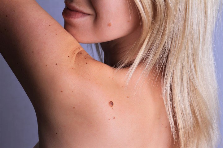
- Location: the dysplastic nevus usually forms in specific areas of the body such as the back, chest, abdomen and extremities. It can also occur in poorly exposed areas such as the breast, scalp, buttocks and groin.
- Evolution: the size of the dysplastic nevus is larger than that of a ‘common’ nevus. Usually a previously stable nevus begins to change, increasing in volume.
- Surface: the central part of the dysplastic nevus is most often raised, while the peripheral area is flat with small elevations.
- Appearance: dysplastic nevi can often assume an unusual shape and are very different from one another.
- Number: a few dysplastic nevi may occur on the body, but also more than a hundred. The number of formations is proportional to the risk of developing melanoma.
Causes
Among the causes that lead to the appearance of a dysplastic nevus are exposure to the sun’s ultraviolet rays without applying sun protection.
Usually those with a lighter phototype are most affected.
Other factors related to dysplastic nevus are ionising radiation.
Dysplastic nevus most often has no symptoms
Itching and discomfort may occur when the lesion appears in specific areas that are subject to anatomical trauma (such as armpits in the groin or skin folds) or trauma from clothing (such as buttons or seams).
Dysplastic nevi are clinically assessed using the ABCDE rule.
“A” – indicates asymmetry: if we ideally divide the lesion into two halves and try to overlap them they do not match.
“B” – the edges of the dysplastic nevus are irregular and map-like.
“C” – the colour is uneven and polychromatic, with different shades of colour in the same lesion.
“D” – indicates the diameter: in dysplastic nevi it is greater than 6 mm.
“E” – indicates evolution and elevation: the first aspect must be investigated through some questions that must be asked to the patient to understand how the lesion has changed over time, the second concerns the relief of the formation with respect to the skin plane.
ABCDE characteristics are not an absolute indication for leading to the removal of moles, particularly when they change over time, but they are useful in requiring periodic follow-up: if they have not changed over time, only periodic (six-monthly or annual) follow-up may be necessary in this case.
On the contrary, the sudden acquisition of such atypical features suggests surgical removal.
Prevention
Prevention, also with regard to dysplastic nevi, remains the best cure for this problem.
In general there are certain rules regarding sun exposure that should be observed by everyone.
First of all, not to expose oneself to the sun during the hottest hours of the day, between 10 a.m. and 4 p.m., is a must.

Artificial lamps and excessive sunbathing should be avoided.
The skin should always be protected from burns and during exposure it is necessary to cover oneself with sunglasses, wide-brimmed hats and clothing.
A UVA and UVB sun filter with a high protection factor should be applied to the skin every day, choosing a product with a water-resistant filter.
The skin should be analysed once a month, and if any suspicious formations appear, it is essential to contact the dermatologist immediately.
A dysplastic nevus is diagnosed during a dermatological examination.
Then the doctor, if he deems it necessary, can perform a biopsy for a histological test.
As already mentioned, it is not always necessary to remove dysplastic moles.
Surgical removal is mainly tested when the moles show initial signs of melanoma or changes that appear after the age of 40.
After the diagnosis of a dysplastic mole is confirmed it is advisable to perform a complete family history indicating atypical moles, melanomas and other tumours in the family.
The patient should also undergo regular check-ups to assess the status of the mole, this visit should be accompanied by a monthly self-examination.
Sun exposure should be reduced, as it may facilitate the formation of new nevi.
Dysplastic nevus and melanoma development
The presence of numerous dysplastic nevi is a risk factor for the development of melanoma.
For this reason it is essential to carefully evaluate the appearance of certain warning signs that could indicate the onset of melanoma.
The most common signs of this disorder are itching, pain, bleeding, elevation, scab formation, swelling, ulceration, blue-blackish colour and the appearance of exudate.
In the presence of these symptoms, it is important to consult a dermatologist immediately.
Scientific studies have shown that individuals with dysplastic moles or a family history of melanoma or dysplastic moles have an increased risk of developing melanoma at an early age.
Melanoma to date is a form of cancer that is easy to identify and therefore curable.
Once diagnosed, the formation must be removed promptly.
If melanoma is left untreated, however, it can progress, giving rise to distant metastases.
It is estimated that around 8,700 people worldwide die of this disease each year.
Especially at an early stage, distinguishing between a melanoma and a dysplastic mole can be difficult.
In this case, the doctor will remove part or all of the lesion and request a histological test to assess its nature.
Another useful tool is the dermatoscope, which allows a magnified view of the nevus and a detailed evaluation of the internal structures that are not visible to the naked eye.
Read Also
Emergency Live Even More…Live: Download The New Free App Of Your Newspaper For IOS And Android
Nevi: What They Are And How To Recognise Melanocytic Moles
Dermatological Examination For Checking Moles: When To Do It
What Is A Tumour And How It Forms
Rare Diseases: New Hope For Erdheim-Chester Disease
How To Recognise And Treat Melanoma
Moles: Knowing Them To Recognise Melanoma
Skin Melanoma: Types, Symptoms, Diagnosis And The Latest Treatments
Melanoma: Prevention And Dermatological Examinations Are Essential Against Skin Cancer
Symptoms And Causes Of Spitz Nevus
What Is A Dysplastic Nevus And What Does It Look Like?
Onychophagia: My Child Bites His Nails, What To Do?
Russia, Doctors Detect Mucormycosis In Covid-19 Patients: What Causes The Fungal Infection?
Parasitology, What Is Schistosomiasis?
Onychomycosis: Why Do Fingernails And Toenails Get Fungus?
Nail Melanoma: Prevention And Early Diagnosis
Ingrown Toenail: What Are The Remedies?
What Is ‘Hand Foot And Mouth’ Disease And How To Recognise It
Dracunculiasis: Transmission, Diagnosis And Treatment Of ‘Guinea-Worm Disease’
Parasitoses And Zoonoses: Echinococcosis And Cystic Hydatidosis
Trichinosis: What It Is, Symptoms, Treatment And How To Prevent Trichinella Infestation
Dermatomycosis: An Overview Of Skin Mycoses