
Epiphysiolysis, an overview of this pathology of the femur
Epiphysiolysis is a condition characterised by the loss of continuity of the femur. In fact, in its upper portion, there is a discontinuity between the head and neck of the bone
Epiphysiolysis is a particular type of epiphyseal detachment and, in some cases, is accompanied by mutual sliding of the two bone portions.
The symptoms of epiphysiolysis can differ depending on the stage of the pathology
The main symptom is pain on movement, which can lead to walking difficulty and make it complex to maintain correct posture.
The combination of these features results in a lame gait.
By making a correct diagnosis, the most appropriate treatment can be set according to the patient’s specific factors.
It should be remembered that the condition, if left untreated, can lead to complications such as instability and hip stiffness.
The ultimate remedy for this condition is surgery.
People who suffer from this condition are mainly adolescents.
In fact, this condition usually affects male patients between the ages of 12 and 17.
The symptoms of this condition can affect one or both femurs, usually 30 to 40 per cent of diagnosed cases are bilateral.
This condition should not be confused with Perthes’ disease which, although it affects the hip in the same way, is a condition that can be classified in the osteochondrosis group.
However, the presenting symptomatology can be confusing and more in-depth investigations may be necessary for a correct differential diagnosis.
Anatomy of the hip
To understand the causes of epiphysiolysis, it is essential to have a basic knowledge of the anatomy of the hip.
This joint is composed of several structures.
The hips are the anatomical region where the union between the trunk and the lower limbs occurs via the coxofemoral joint.
It is composed of the juxtaposition of the cup of the iliac bone with the head of the femur.
Numerous structures such as tendons, ligaments and muscles are also present in this anatomical region.
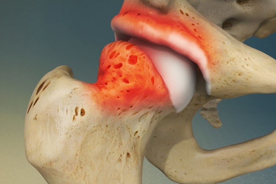
The epiphysis at the rounded end of the long bones is connected to the diaphysis through the metaphyses.
Epiphysiolysis is an alteration that results in a rupture of the epiphysis after which the normal relationship between the head and neck of the femur is lost.
The distal component may slip relative to the proximal component, worsening the clinical picture.
This condition is also known as SCFE, Slipped Capital Femoral Epiphysis.
The growth and consolidation of tissue and bone in adolescence can be altered in the presence of different risk factors.
These include trauma, genetic predisposition and being overweight.
Depending on the severity, two different clinical entities are recognised:
- stable epiphysiolysis, which is characterised by milder symptoms and the patient is able to perform simple actions;
- unstable epiphysiolysis, which is the most severe form and is characterised by the patient being unable to walk or put his weight on his hip.
To avoid complications, treatment must be timely.
The symptoms of epiphysiolysis differ depending on the stage of severity of the patient’s pathology
The cardinal symptom of the pathological condition is pain.
This may present acutely or slowly emerge into chronic.
Pain, at least initially, occurs during movement or sporting activity.
Painful symptoms can make walking complicated and alter the posture of the subject, who tries to maintain an antalgic attitude.
Because of the pain, it is common for the patient to limp, as he or she tends to put all the weight of the body on the non-painful lower limb.
This can also lead to back problems in the long term.
Several signs may emerge during the objective test:
- difficulty in internal rotation of the hip;
- inability to flex the hip;
- pain in hip abduction;
- decreased range of motion.
In addition to these symptoms, there may also be muscle stiffness.
The pain may also extend to other parts of the body such as the groin, knee and thigh.
This non-specific symptomatology is insidious as it can delay the diagnosis of the condition.
The causes of epiphysiolysis are still not totally clear
In fact, it is not possible to attribute the onset of the pathology to a single factor.
Nevertheless, it is possible to recognise the existence of certain risk factors that may predispose to epiphysiolysis.
Remembering that the pathology affects the growth nucleus of the femoral head before it has reached complete consolidation, age is not a risk factor.
Amongst this we recognise:
- Body weight above normal: in children with childhood obesity, the likelihood of contracting this pathology increases; data show that a large proportion of patients with this pathology are above the 95th percentile for body weight.
- Genetic predisposition: it is not uncommon for first-degree relatives of an affected person to have other cases of the same disease.
- Hormonal factors: certain hormonal alterations can disrupt the correct development of the femoral epiphysis, predisposing to the disease, the hormones involved being sexual or thyroid hormones.
Diagnosis of epiphysiolysis is not very complex
Despite the poor specificity of the symptoms, a few simple tests are sufficient to confirm the diagnosis.
During the first visit the patient will report his or her symptoms with particular attention to the type of pain perceived, the mode of onset and the response to any pharmacological treatment.
The patient should then be weighed and this data should be compared with the reference value for his or her age.
Thanks to this information, the doctor will be able to raise a diagnostic suspicion, which will have to be confirmed by performing some specific tests.

Following this interview, the practitioner may prescribe, if deemed necessary, a visit with a specialist, usually an orthopaedist.
At the specialist’s discretion, further investigations may be required:
- X-ray tests, which can provide a detailed picture of the bone structure and condition of the hip. X-rays of the pelvis, thigh and hip are usually requested;
- In selected cases, an MRI scan may be necessary.
This allows better visualisation of the surrounding soft tissues.
Treatment can be conservative or surgical
The former are recommended in cases of mild pathology.
The main conservative treatment is rest, the patient should refrain from sporting activity and, in severe cases, should avoid walking without adequate support.
If the pathology does not resolve with these measures, or if it already appears with important alterations of the anatomical structures, surgery will be necessary.
The aim of the operation is to stabilise the femoral head and prevent complications.
The operation is essential in order to avoid the risk of developing early arthrosis of the hip and, in addition, provides rapid relief from pain symptoms.
Following the operation, several days of monitoring in hospital and a rest period determined by the surgeon will be necessary.
Coupled with the surgery, a course of physiotherapy may be set up to help regain correct and complete mobility in the joint.
In addition, these physiotherapy sessions can lead to increased muscle strength and greater stability of the lumbopelvic complex.
Most patients who are diagnosed with epiphysiolysis are at a developing age, which is why it is essential to carry out periodic checks after the operation.
In this way, the patient’s growth status and recovery can be analysed.
Epiphyseolysis of the femur must be diagnosed quickly in order to prevent possible complications
These can include necrosis, which can occur when the femoral epiphyseolysis is very severe or is neglected. In this case, the blood supply to the femoral head is reduced.
This causes significant pain in the hip and may make it necessary to take pain-relieving medication on a chronic basis.
Another very rare complication of epiphysiolysis is chondrolysis.
This condition leads to the degeneration of the cells that make up the articular cartilage and their replacement by bone tissue.
Read Also
Emergency Live Even More…Live: Download The New Free App Of Your Newspaper For IOS And Android
Hip Pain: Causes, Symptoms, Diagnosis, Complications, And Treatment
Cartilage Injuries: Chondropathy
MOP Hip Implant: What Is It And What Are The Advantages Of Metal On Polyethylene
Hip Osteoarthritis: What Is Coxarthrosis
Why It Comes And How To Relieve Hip Pain
Hip Arthritis In The Young: Cartilage Degeneration Of The Coxofemoral Joint
Visualizing Pain: Injuries From Whiplash Made Visible With New Scanning Approach
Coxalgia: What Is It And What Is The Surgery To Resolve Hip Pain?
Elbow Fracture: What To Do After A Fall And Healing Time
Treating Injuries: When Do I Need A Knee Brace?
Wrist Fracture: How To Recognise And Treat It
Carpal Tunnel Syndrome: Diagnosis And Treatment
How To Put On Elbow And Knee Bandages
Knee Ligament Rupture: Symptoms And Causes
Lateral Knee Pain? Could Be Iliotibial Band Syndrome
Knee Sprains And Meniscal Injuries: How To Treat Them?
Stress Fractures: Risk Factors And Symptoms
What Is OCD (Obsessive Compulsive Disorder)?
RICE Treatment For Soft Tissue Injuries
P.O.L.I.C.E. Vs R.I.C.E.: The Emergency Treatment For Acute Injuries
How And When To Use A Tourniquet: Instructions For Creating And Using A Tourniquet
Bone Callus And Pseudoarthrosis, When The Fracture Does Not Heal: Causes, Diagnosis And Treatment
First Aid, Fractures (Broken Bones): Find Out What To Look For And What To Do
Epicondylitis Or Tennis Elbow: How To Treat It?
Golfer’s Elbow, An Overview Of Epitrocleitis