
In-depth analysis of the heart: Cardiac Magnetic Resonance Imaging (CARDIO - MRI)
Cardiac Magnetic Resonance Imaging (CARDIO – MRI) represents one of the latest and most innovative diagnostic methods in the field of cardiac
Cardiac Magnetic Resonance Imaging (CARDIO – MRI), what are its main applications?
- Precise and reliable assessment of ventricular volumes, ventricular ejection fraction and myocardial mass, using techniques already in use in echocardiography, but with greater definition of the endocardial and epicardial contours, so much so that it can now be considered the gold standard method due to its excellent reproducibility;
- Study of myocardial perfusion and contractile reserve by combining in a single diagnostic method the information generally obtained from different tests such as echo-stress and nuclear medicine tests;
- Study of myocardial viability by detecting scarring areas of the myocardial wall, now fundamental both in the indication of revascularisation procedures and in the prognostic stratification of patients in the most varied heart diseases.
FIRST AID: VISIT THE DMC DINAS MEDICAL CONSULTANTS BOOTH AT EMERGENCY EXPO
Thanks to this triple assessment, CARDIO – MRI can be used to investigate countless clinical contexts
These include ischaemic heart disease, dilated cardiomyopathy, myocarditis, hypertrophic cardiomyopathy, arrhythmogenic right ventricle disease, congenital heart disease, valvulopathies, pericardial disease and the study of cardiac masses.
MRI of the heart is a second-level test for evaluating most heart and heart valve diseases
In detail, MRI of the heart is a magnetic resonance imaging (MRI) that allows the heart muscle in motion to be analysed by acquiring images that are then rearranged into a dynamic video.
Thanks to this technique, it is possible to study the anatomy and functioning of the heart, atria, great vessels and heart valves, thus intercepting possible diseases, both congenital and acquired.
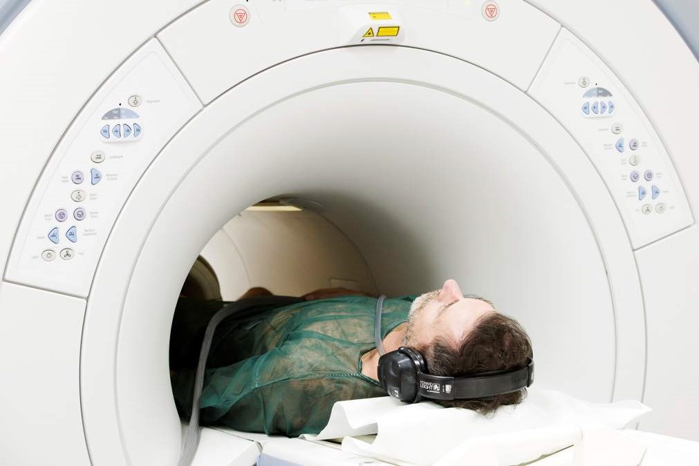
As far as the execution of the test is concerned, firstly, the patient is made to lie on a couch, where the electrodes for monitoring the heartbeat and the surface coils necessary for the analysis are applied.
For the MRI scan to be successful, it is essential that the patient is able to hold his or her breath for at least ten seconds, as the scans are performed in apnoea and with heartbeat monitoring, in order to eliminate artefacts from cardiac and respiratory motion.
In most cases, a contrast medium will be administered intravenously, about halfway through the test, to allow, for example, a diagnostic assessment of possible structural damage of the myocardium as a result of ischaemic or inflammatory events.
In addition, defects in myocardial perfusion can also be detected during the administration of the mdc in the early phase.
The duration of the test varies from case to case, with a range of 45-60 minutes.
TOP AMBULANCES AND MEDICAL INTERVENTION EQUIPMENT? VISIT THE DIAC MEDICAL BOOTH AT EMERGENCY EXPO
Read Also
Emergency Live Even More…Live: Download The New Free App Of Your Newspaper For IOS And Android

Nuclear Magnetic Resonance (NMR): When To Do It?
Heart Attack: Guidelines For Recognising Symptoms
Medical Thermography: What Is It For?
Rheumatic Diseases: The Role Of Total Body MRI In Diagnosis
Positron Emission Tomography (PET): What It Is, How It Works And What It Is Used For
Single Photon Emission Computed Tomography (SPECT): What It Is And When To Perform It
Instrumental Examinations: What Is The Colour Doppler Echocardiogram?
Coronarography, What Is This Examination?
CT, MRI And PET Scans: What Are They For?
MRI, Magnetic Resonance Imaging Of The Heart: What Is It And Why Is It Important?
Urethrocistoscopy: What It Is And How Transurethral Cystoscopy Is Performed
What Is Echocolordoppler Of The Supra-Aortic Trunks (Carotids)?
Surgery: Neuronavigation And Monitoring Of Brain Function
Robotic Surgery: Benefits And Risks
Refractive Surgery: What Is It For, How Is It Performed And What To Do?
What Is Magnetic Resonance Angiography (MRA)?