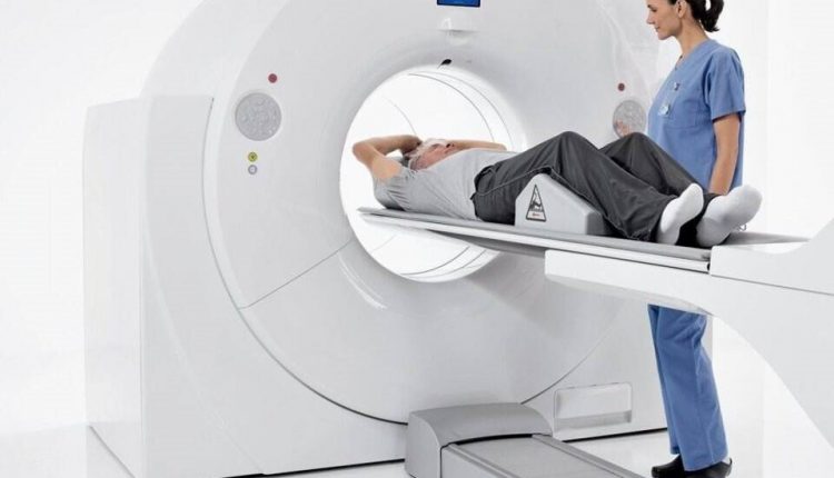
Positron Emission Tomography (PET): what it is, how it works and what it is used for
Positron Emission Tomography (PET) is an innovative instrumental investigation that uses radioactive compounds to diagnose abnormalities at the molecular biological level that often precede anatomical alteration
How PET works
PET is based on the introduction into the patient’s body (usually intravenously) of radioactive compounds (molecular probes or ‘tracers’) chemically bound to a metabolically active molecule (often a sugar) and which have a different rate of absorption depending on the tissue involved.
As soon as the metabolically active molecule reaches a certain concentration within the organic tissue to be analysed, the subject is placed in the scanner.
When the radioactive compound ‘decays’, it releases positrons (positive electrons), which in turn release gamma electromagnetic radiation: this is recorded by the scanner and processed electronically to compose an image capable of visualising the regions where there are accumulations of the molecular probe.
The resulting map is read and interpreted by a specialist in nuclear medicine or radiology in order to determine a diagnosis and subsequent treatment.
What PET is used for
PET scanning is used in oncology to obtain images of tumours, to search for metastases and to monitor the response to anti-cancer therapies.
In neurology it is used to visualise brain regions with a particularly active metabolism because they are affected by disease (as in the case of epileptic outbreaks) or to highlight a loss of brain activity.
In cardiology, it is used above all to study myocardial blood supply: with a PET scan, it is possible to visualise and quantify the change in blood flow in the various anatomical structures with fair accuracy.
Read Also
Emergency Live Even More…Live: Download The New Free App Of Your Newspaper For IOS And Android
Single Photon Emission Computed Tomography (SPECT): What It Is And When To Perform It
Instrumental Examinations: What Is The Colour Doppler Echocardiogram?
Coronarography, What Is This Examination?
CT, MRI And PET Scans: What Are They For?
MRI, Magnetic Resonance Imaging Of The Heart: What Is It And Why Is It Important?
Urethrocistoscopy: What It Is And How Transurethral Cystoscopy Is Performed
What Is Echocolordoppler Of The Supra-Aortic Trunks (Carotids)?
Surgery: Neuronavigation And Monitoring Of Brain Function
Robotic Surgery: Benefits And Risks
Refractive Surgery: What Is It For, How Is It Performed And What To Do?


