
Instrumental examinations: what is the colour Doppler echocardiogram?
An echocardiogram is an instrumental examination that enables a morphological and functional assessment of the heart
There are different types of echocardiogram:
- the transthoracic (or standard) echocardiogram;
- the transesophageal echocardiogram, provides more precise images of the heart and associated blood vessels, however it is characterised by greater invasiveness as the probe is inserted through the patient’s oesophagus;
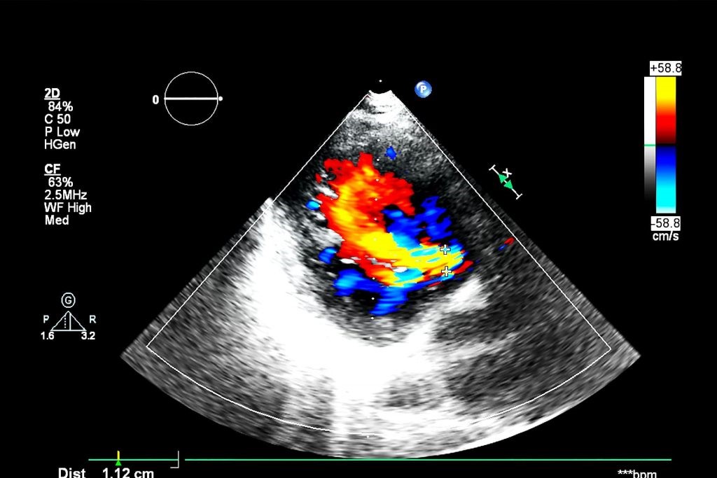
- the colour-Doppler echocardiogram, which can be performed either in transthoracic or transesophageal mode as required. It makes it possible to highlight blood flow with greater precision thanks to colour-Doppler technology;
- the stress echocardiogram, performed following intense physical activity, is used to assess cardiac behaviour when the heart is subjected to stress.
These examinations are performed using a special instrument called an echocardiograph.
During the measurement, a small ultrasound probe is placed gently on the patient’s chest, returning images of the anatomical structures of the heart on a screen; thanks to an echocardiogram, it is then possible to accurately analyse the health of the heart, the condition of the heart walls, the valve leaflets, etc.
At the same time, the echocardiogram recording involves studying the blood flow within the heart chambers (i.e. the atria and ventricles) and the valve apparatus.
In particular, the colour Doppler echocardiogram highlights blood flows marked by two different colours: red for flows moving in the direction of the probe (and therefore the heart), and blue for flows moving away from it.
The echocardiogram makes it possible to study the cardiac anatomy, as well as the mechanical functions of the heart
Thanks to this assessment, it is also possible to evaluate the contractile power of the heart muscle in order to detect any alterations.
This examination is very important for screening and monitoring heart disease over time.
Finally, an echocardiogram can be useful for assessing the outcomes of cardiac surgery and drug therapies.
The colour Doppler echocardiogram, or simply echocolordoppler, is a special type of cardiac ultrasound, used to
- detect possible damage to the myocardium
- establish the damage of a myocardial infarction or ischaemia;
- diagnose heart failure;
- examine congenital heart defects;
- identify abnormalities in heart valves or valvulopathies;
- diagnose cardiomyopathies;
- determine the presence of infections and inflammations such as endocarditis, pericarditis or myocarditis.
How should I prepare?
The echocardiogram is a non-invasive examination that is carried out in the same way as a normal ultrasound scan: the patient is laid on a couch in a supine position or on their side, after which a thin layer of a special gel that conducts the waves is smeared on them.
No special preparation is then required.
Ultrasound, in fact, is nothing more than sound waves that are inaudible to the human ear: it does not involve exposure to radiation and therefore the echocardiogram is a harmless examination with no side effects; it can be repeated several times and has no contraindications for pregnant women, children or the elderly.
*This is only indicative information: it is therefore necessary to contact the facility where the examination is being performed to obtain specific information on the preparation procedure.
Read Also
Emergency Live Even More…Live: Download The New Free App Of Your Newspaper For IOS And Android
Identifying Tachycardias: What It Is, What It Causes And How To Intervene On A Tachycardia
Tachycardia: Is There A Risk Of Arrhythmia? What Differences Exist Between The Two?
Do You Have Episodes Of Sudden Tachycardia? You May Suffer From Wolff-Parkinson-White Syndrome (WPW)
Transient Tachypnoea Of The Newborn: Overview Of Neonatal Wet Lung Syndrome
Paediatric Toxicological Emergencies: Medical Intervention In Cases Of Paediatric Poisoning

Valvulopathies: Examining Heart Valve Problems
What Is The Difference Between Pacemaker And Subcutaneous Defibrillator?
Heart Disease: What Is Cardiomyopathy?
Inflammations Of The Heart: Myocarditis, Infective Endocarditis And Pericarditis
Heart Murmurs: What It Is And When To Be Concerned
Broken Heart Syndrome Is On The Rise: We Know Takotsubo Cardiomyopathy
Cardiomyopathies: What They Are And What Are The Treatments
Alcoholic And Arrhythmogenic Right Ventricular Cardiomyopathy
Difference Between Spontaneous, Electrical And Pharmacological Cardioversion
What Is Takotsubo Cardiomyopathy (Broken Heart Syndrome)?
Dilated Cardiomyopathy: What It Is, What Causes It And How It Is Treated
Heart Pacemaker: How Does It Work?
Basic Airway Assessment: An Overview
Assessment Of Abdominal Trauma: Inspection, Auscultation And Palpation Of The Patient
Pain Assessment: Which Parameters And Scales To Use When Rescuing And Treating A Patient
Airway Management After A Road Accident: An Overview
Tracheal Intubation: When, How And Why To Create An Artificial Airway For The Patient
What Is Traumatic Brain Injury (TBI)?
Acute Abdomen: Meaning, History, Diagnosis And Treatment
Poison Mushroom Poisoning: What To Do? How Does Poisoning Manifest Itself?
Chest Trauma: Clinical Aspects, Therapy, Airway And Ventilatory Assistance
The Quick And Dirty Guide To Pediatric Assessment
EMS: Pediatric SVT (Supraventricular Tachycardia) Vs Sinus Tachycardia
Supraventricular Tachycardia: Definition, Diagnosis, Treatment, And Prognosis