
Myocardial scintigraphy, the examination that describes the health of the coronary arteries and myocardium
Myocardial scintigraphy is a diagnostic examination to examine the health of the coronary arteries and the blood supply to the myocardium
Myocardial scintigraphy, what is it?
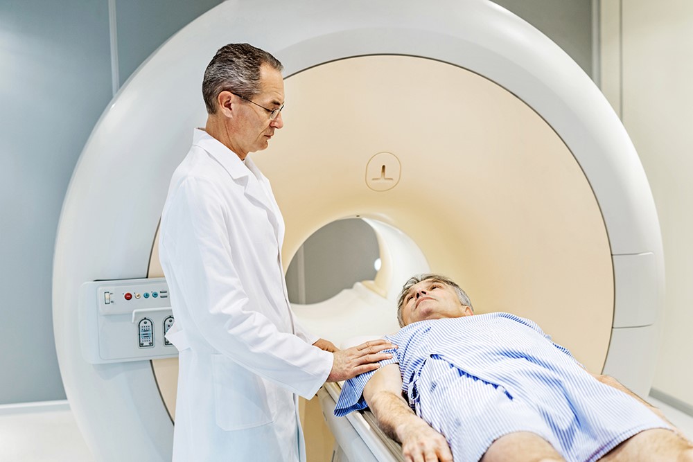
This is a nuclear medicine technique, which involves the injection of radiopharmaceuticals (i.e. substances containing radioactive isotopes or radionuclides), which are tracked with a special detection instrument, providing very detailed images of the body.
Scintigraphy techniques are used to investigate the location, shape, size or function of various organs and anatomical structures, including the heart, thyroid, bones, brain, liver, kidneys or lungs.
Once injected, radiopharmaceuticals interact specifically with certain types of biological tissues; thanks to their radioactive properties, it is possible to examine their diffusion through a special gamma camera, which returns very clear and meaningful images.
Myocardial scintigraphy makes it possible to carefully analyse the propagation of blood flow within the coronary arteries, the perfusion of the myocardium and the function of the heart.
The procedure involves 2 stages: first, the heart is analysed while the patient is under physical exertion; then, after an appropriate interval, the procedure is repeated with the patient at rest.
In some cases, exercise myocardial scintigraphy can be replaced by pharmacological stress myocardial scintigraphy, in which drugs are administered to simulate cardiac behaviour during physical activity. In this way, the cardiologist is able to compare myocardial blood flow during stress conditions and at rest.
Why myocardial scintigraphy is performed
Generally, myocardial scintigraphy is performed when the presence of coronary artery disease, damage to the coronary arteries or narrowing of the blood vessels responsible for perfusing the myocardium is suspected; the main causes of stenosis or occlusion of the coronary arteries may be blood clots, or so-called atheromasic plaques (i.e. deposits of lipids, platelets, white blood cells and muscle cells).
If the narrowing is greater than 70 per cent, the blood supply may be insufficient to support cardiac activity, leading to a condition of coronary ischaemia which, if not adequately treated, can lead to a myocardial infarction.
Scintigraphy is also often used to analyse the damage of a heart attack and identify the necrotic portion of myocardium; or to assess the outcome of therapeutic treatments to restore coronary flow, such as bypass and angioplasty with stenting.
How should I prepare for myocardial scintigraphy
Myocardial scintigraphy is a non-invasive examination that requires special preparation; prior to the procedure, the doctor subjects the patient to a thorough objective examination, during which he will explain all the indications necessary for the procedure and assess the presence of any contraindications.
On the day of the examination, the patient is required to fast completely for at least 12 hours and, depending on the case, it may be necessary to interrupt any pharmacological therapies.
The cardiologist should also be informed of any special medical conditions or if you have arrhythmia correction devices.
*This is only indicative information: it is therefore necessary to contact the facility where the examination is being performed to obtain specific information on the preparation procedure.
Read Also
Head Up Tilt Test, How The Test That Investigates The Causes Of Vagal Syncope Works
Aslanger Pattern: Another OMI?
Abdominal Aortic Aneurysm: Epidemiology And Diagnosis
What Is Ischaemic Heart Disease And Possible Treatments
Percutaneous Transluminal Coronary Angioplasty (PTCA): What Is It?
Ischaemic Heart Disease: What Is It?
EMS: Pediatric SVT (Supraventricular Tachycardia) Vs Sinus Tachycardia
Paediatric Toxicological Emergencies: Medical Intervention In Cases Of Paediatric Poisoning
Valvulopathies: Examining Heart Valve Problems

What Is The Difference Between Pacemaker And Subcutaneous Defibrillator?
Heart Disease: What Is Cardiomyopathy?
Inflammations Of The Heart: Myocarditis, Infective Endocarditis And Pericarditis
Heart Murmurs: What It Is And When To Be Concerned
Clinical Review: Acute Respiratory Distress Syndrome
Botallo’s Ductus Arteriosus: Interventional Therapy
Heart Valve Diseases: An Overview
Cardiomyopathies: Types, Diagnosis And Treatment
First Aid And Emergency Interventions: Syncope
Tilt Test: What Does This Test Consist Of?
Cardiac Syncope: What It Is, How It Is Diagnosed And Who It Affects
New Epilepsy Warning Device Could Save Thousands Of Lives
Understanding Seizures And Epilepsy
First Aid And Epilepsy: How To Recognise A Seizure And Help A Patient
Neurology, Difference Between Epilepsy And Syncope
Positive And Negative Lasègue Sign In Semeiotics
Wasserman’s Sign (Inverse Lasègue) Positive In Semeiotics
Positive And Negative Kernig’s Sign: Semeiotics In Meningitis
Lithotomy Position: What It Is, When It Is Used And What Advantages It Brings To Patient Care
Trendelenburg (Anti-Shock) Position: What It Is And When It Is Recommended
Prone, Supine, Lateral Decubitus: Meaning, Position And Injuries
Stretchers In The UK: Which Are The Most Used?
Does The Recovery Position In First Aid Actually Work?
Reverse Trendelenburg Position: What It Is And When It Is Recommended
Drug Therapy For Typical Arrhythmias In Emergency Patients
Canadian Syncope Risk Score – In Case Of Syncope, Patients Are Really In Danger Or Not?