
Patellar luxation: causes, symptoms, diagnosis and treatment
Patellar luxation is the dislocation of the kneecap. Different and less serious than knee luxation, it consists of the displacement of the kneecap, which goes out of place
This usually occurs as a result of an abrupt change in direction and movement and occurs quite often in adolescents, especially if they are sporty.
It is a temporary dislocation, which can be put back in place by a doctor, to whom one must necessarily turn in the event of a dislocation.
In some cases (the less serious ones), the kneecap goes back in place on its own, in others the doctor’s manual intervention is needed.
In still others, and this is where the more serious ones come in, surgery is necessary.
It should be specified that luxating the kneecap is less serious than luxating the knee.
The kneecap is in fact a small protective bone that attaches near the lower part of the femur.
It moves up and down in a groove called the trochlea, allowing the knee to bend and straighten.
The kneecap is held in place by muscles and ligaments, which, in the event of an injury, can cause pain and difficulty bending the knee.
Depending on the severity of the injury, the doctor will determine whether what he or she has in front of him or her is a subluxation or a true dislocation.
In most injuries, the kneecap is pushed outwards.
This can also damage the medial patellofemoral ligament, which, if it does not heal properly, can set the stage for a second dislocation.
What are the different degrees of severity
Depending on the severity of the injury, there will either be a patellar subluxation or a patellar dislocation.
The former is a common injury in children and adolescents, but can also occur in adulthood.
It is a non-serious event that normally does not require surgery.
However, in the case of a more serious injury requiring surgery, new intervention techniques are now so advanced that a full recovery is very likely.
In the case of more serious injuries, it could happen that the knee ligament, the medial patellofemoral ligament, is injured.
Should this occur, it is a good idea to follow the doctor’s instructions to the letter to ensure a full recovery.
If this does not happen, and recovery is not 100 per cent, it is very likely that a second patellar luxation will occur after a short time.
Causes of patellar luxation
Most first injuries occur during sport.
Abrupt changes of direction, such as in football, gymnastics, contact sports, and basketball, can cause the patella to shift and dislocate.
It is no coincidence that injuries mainly affect young, active people.
Symptoms and complications of patellar luxation
One is usually immediately aware when the patella goes out of place.
This is because it often occurs following an abrupt movement, and is also quite visible, depending on the severity of the injury.
However, there are several common symptoms:
- instability of the knee and sliding of the kneecap outwards;
- pain after prolonged movement;
- pain in the front part of the knee that worsens markedly after physical activity;
- stiffness and swelling of the knee.
As mentioned, it is not difficult to realise that one’s kneecap is dislocated.
In any case, even if the diagnosis has been made independently, it is always necessary to see a doctor for treatment.
You could be faced with a simple dislocation, which can go away on its own or with a simple manoeuvre, or a severe dislocation requiring surgery.
The most serious complication, in the case of a very serious injury, is injury to the knee ligaments.
Patellar luxation diagnosis
To diagnose a patellar luxation, the doctor will bend and straighten the injured knee, feeling the area around the kneecap.
Once the injury is established, the doctor may decide to proceed with X-rays.
An MRI scan will be used to visualise the ligaments and other soft tissues around the kneecap.
Therapy
In cases of patellar luxation, there are two options: cases that do not require surgery, and those that do.
Non-surgical methods
Most patellar luxations do not require surgical treatment.
In the case of a subluxation or a first injury, a different procedure is always attempted, with the doctor gently moving the patella back into place.
In this case, the luxation is treated by putting the person at rest and having them do specific exercises to put the kneecap back in place.
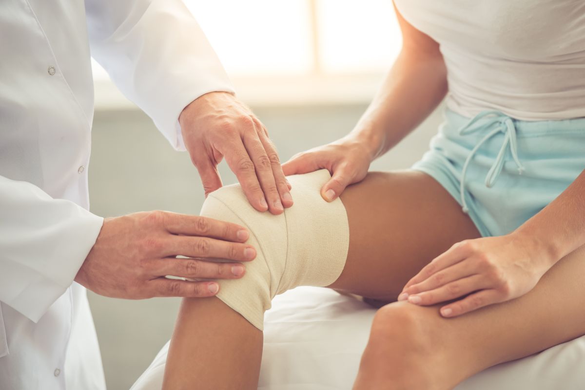
The doctor may also prescribe a course of physiotherapy to fully recover, and the use of a crutch to take weight off the knee.
As well as braces to immobilise it and specialised footwear to relieve pressure on the kneecap.
The chance of recurrence is just over 30%.
Surgical methods
In most cases, an initial patellar luxation is treated with non-surgical methods.
Operations are only recommended on special occasions, and in the case of repeated episodes.
One of the operations that one can undergo is the reconstruction of the medial patellofemoral ligament.
This is the ligament that pulls the kneecap towards the inside of the leg.
When this is weak or damaged, the kneecap can dislocate outwards.

In this operation, the ligament is reconstructed using a small piece of tendon taken either from a donor or from the biceps femoris muscle.
These operations are generally performed in an outpatient setting: once you leave the clinic or hospital, a brace is applied to stabilise the knee.
The task of the brace is to keep the leg straight while walking and is worn – depending on the severity of the case – for one to two months.
At the end of this period, rehabilitation begins, which usually consists of physiotherapy.
The recovery time is not very short; most people can resume sport even after seven months after the injury.
Another type of operation is that involving the transfer of the tibial tuberosity.
An injury that has caused dislocation of the kneecap may have damaged the connection point with this tendon.
In this case, the doctor has to transfer a small piece of the tibial tuberosity to improve the attachment of the tendon and help the patella move correctly in its scaling.
After the operation, the patient will have to use crutches to walk for as long as indicated by the doctor.
Afterwards, he/she will begin with physiotherapy.
Patellar luxation prevention
Certain exercises can help strengthen the leg muscles and reduce the possibility of injury not only to the kneecap, but also to the knee (an even more serious case that is best avoided).
These are exercises that can be done every day and take little time.
Even if you find them tedious to do, it is good to perform them to strengthen the muscles and decrease the likelihood of patellar luxation.
Some of the exercises that can be done to prevent patellar luxation are squats and leg lifts.
In the case of individuals who have already suffered a patellar luxation, wearing a brace can help prevent recurrences.
Another way to prevent dislocations is to wear appropriate protective clothing in contact sports, so as to protect the knee in the event of blows that can injure the leg and knee.
Read Also
Emergency Live Even More…Live: Download The New Free App Of Your Newspaper For IOS And Android
Work-Related Musculoskeletal Disorders: We Can All Be Affected
Arthrosis Of The Knee: An Overview Of Gonarthrosis
Varus Knee: What Is It And How Is It Treated?
Patellar Chondropathy: Definition, Symptoms, Causes, Diagnosis And Treatment Of Jumper’s Knee
Jumping Knee: Symptoms, Diagnosis And Treatment Of Patellar Tendinopathy
Symptoms And Causes Of Patella Chondropathy
Unicompartmental Prosthesis: The Answer To Gonarthrosis
Anterior Cruciate Ligament Injury: Symptoms, Diagnosis And Treatment
Ligaments Injuries: Symptoms, Diagnosis And Treatment
Knee Arthrosis (Gonarthrosis): The Various Types Of ‘Customised’ Prosthesis
Rotator Cuff Injuries: New Minimally Invasive Therapies
Knee Ligament Rupture: Symptoms And Causes
MOP Hip Implant: What Is It And What Are The Advantages Of Metal On Polyethylene
Hip Pain: Causes, Symptoms, Diagnosis, Complications, And Treatment
Hip Osteoarthritis: What Is Coxarthrosis
Why It Comes And How To Relieve Hip Pain
Hip Arthritis In The Young: Cartilage Degeneration Of The Coxofemoral Joint
Visualizing Pain: Injuries From Whiplash Made Visible With New Scanning Approach
Coxalgia: What Is It And What Is The Surgery To Resolve Hip Pain?
Lumbago: What It Is And How To Treat It
Lumbar Puncture: What Is A LP?
General Or Local A.? Discover The Different Types
Intubation Under A.: How Does It Work?
How Does Loco-Regional Anaesthesia Work?
Are Anaesthesiologists Fundamental For Air Ambulance Medicine?
Epidural For Pain Relief After Surgery
Lumbar Puncture: What Is A Spinal Tap?
Lumbar Puncture (Spinal Tap): What It Consists Of, What It Is Used For
What Is Lumbar Stenosis And How To Treat It
Lumbar Spinal Stenosis: Definition, Causes, Symptoms, Diagnosis And Treatment