
Abdominal ultrasound: how it is performed and what it is used for
Abdominal ultrasound is an investigation that allows the doctor to explore the abdominal organs
It uses ultrasound, which is emitted by a probe resting on the skin that also picks up their reflection, which varies according to the different consistency of the tissues passed through.
With the addition of the Doppler, it is possible to obtain information on the circulation within the vessels and the vascularisation of any masses.
The Doppler effect can be represented on the screen either by graphic and sound signals or by a ‘colour effect’ within the vessels (colour Doppler).
Today it is also possible, with the use of special contrast media, to further enhance the capabilities of the Doppler by improving its sensitivity.
When to perform abdominal ultrasound
Ultrasound is primarily used to assess the shape, size and structure of organs.
Through this information, pathologies of various kinds affecting all abdominal organs can be diagnosed.
In particular, it is used to assess
- acute and chronic liver diseases (hepatitis, cirrhosis, etc.);
- diseases of the gallbladder and biliary tract (gallstones and inflammation of the gallbladder, biliary obstruction, etc.)
- pancreatic diseases (pancreatitis, solid or cystic neoformations);
- kidney diseases (acute and chronic nephritis, stones, urinary tract obstructions, etc.);
- diseases of the spleen and abdominal lymph nodes (increased volume);
- space-occupying masses and lesions (benign and malignant tumours, cysts, abscesses, etc.);
- presence of free fluid or collections in the abdominal cavity;
- changes in venous and arterial vessels (aortic aneurysm, increased calibre of the portal vein, etc.);
- changes in the thickness of the intestinal wall (chronic inflammatory diseases) or dilation of intestinal segments (as a result of obstructions).
With the aid of ultrasound it is possible to carry out minor operations using special needles and catheters to treat tumours (by injecting substances into them or inserting thermal probes or lasers) or take tissue samples (biopsies).
Abdominal ultrasound is not used to assess changes in the intestinal mucosa; if there is a lot of abdominal meteorism, it can be difficult to assess certain organs, especially the pancreas, in obese people.
How abdominal ultrasound is performed
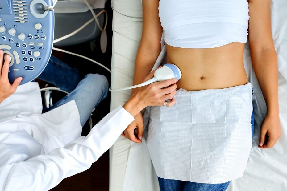
It is performed by placing the ultrasound probe on the abdomen.
To improve ultrasound transmission, a watery gel is placed between the probe and the skin.
No special precautions are necessary; it may be useful to practise a diet without fruit and vegetables in the preceding days to reduce intestinal meteorism.
It can be performed in any environment (even at home).
How to prepare for abdominal ultrasound
The test is performed with the patient having been fasting for at least six hours. As far as is known today, ultrasound is harmless.
The test generally lasts only a few minutes and is absolutely painless and does not cause any discomfort.
It is performed with the patient lying supine and/or on their side.
Read Also
Emergency Live Even More…Live: Download The New Free App Of Your Newspaper For IOS And Android
Abdominal Pain Emergencies: How US Rescuers Intervene
Abdominal Distension (Distended Abdomen): What It Is And What It Is Caused By
Umbilical Cord: What Is It, What Is It For, What Does It Contain?

Abdominoplasty (Tummy Tuck): What It Is And When It Is Performed
Assessment Of Abdominal Trauma: Inspection, Auscultation And Palpation Of The Patient
What’s Causing Your Abdominal Pain And How To Treat It
Intestinal Infections: How Is Dientamoeba Fragilis Infection Contracted?
Early Parenteral Nutrition Support Cuts Infections After Major Abdominal Surgery
Acute Abdomen: Meaning, History, Diagnosis And Treatment
Abdominal Trauma: A General Overview Of Management And Trauma Areas
Fusion Prostate Biopsy: How The Examination Is Performed
What Is Needle Aspiration (Or Needle Biopsy Or Biopsy)?
What Is Echocolordoppler Of The Supra-Aortic Trunks (Carotids)?
What Is The Loop Recorder? Discovering Home Telemetry
Cardiac Holter, The Characteristics Of The 24-Hour Electrocardiogram
Peripheral Arteriopathy: Symptoms And Diagnosis
Endocavitary Electrophysiological Study: What Does This Examination Consist Of?
Cardiac Catheterisation, What Is This Examination?
Echo Doppler: What It Is And What It Is For
Transesophageal Echocardiogram: What Does It Consist Of?
Venous Thrombosis: From Symptoms To New Drugs
Echotomography Of Carotid Axes
Echo- And CT-Guided Biopsy: What It Is And When It Is Needed
Echodoppler: What It Is And When To Perform It