
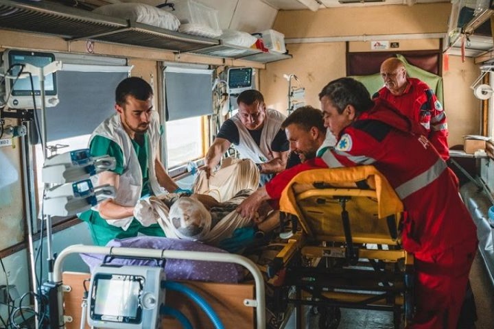
Chest trauma: symptoms, diagnosis and management of the patient with severe chest injury
A person will be diagnosed with chest trauma when they have a severe chest injury
Also known as chest trauma, this condition will cause disability and death may occur as a consequence; it is the third leading cause of death from physical trauma.
Chest trauma may occur as a result of a variety of injuries; among the most common causes of chest injuries are traffic accidents.
Accidental or malicious injuries can cause thoracic trauma
Chest injuries include gunshot wounds, can also occur as a result of falls, after being stabbed, hit or beaten.
A diagnosis can be made by a physician, usually with an X-ray.
Of course, thoracic trauma is one of the most complex topics, and in the in-depth articles it will be possible to learn about other peculiar aspects: it is impossible to summarise the subject in one text.
Chest trauma can be divided into two types:
- Penetrating trauma, which occurs when the victim suffers an injury that breaks the skin, such as a knife in the chest or a gunshot wound;
- Bruising trauma will result in some tearing of the skin, the tear is not the cause of the injury itself and the damage is often less localised. Being kicked by a large animal or being in a car accident can cause blunt trauma.
Blunt trauma accounts for 25% of all deaths due to traumatic medical emergencies.
Chest trauma will present several symptoms, the most common of which will be intense pain and difficulty breathing.
Other symptoms will include bleeding, shock, shortness of breath, bleeding, bruising and loss of consciousness, which will occur depending on the cause of the chest trauma.
Bone fracture may also occur due to a thoracic injury.
Chest trauma will be treated depending on the cause
Interventions may be required to clear the airway, both in case of lung collapse and to prevent the trauma from causing worse damage and thus resulting in infection.
Trauma to the chest can cause various forms of cardiac injury, such as penetration of a foreign body, rupture, tamponade, laceration and occlusion of the coronary arteries, myocardial contusion, pericardial effusion, septal defects, valvular lesions, and rupture of the large vessels.
These injuries are often fatal.
Penetrating cardiac injuries are most often caused by blunt weapons or shotguns and result in a mortality rate of between 50% and 85%.
Closed traumas are most frequently associated with a rupture of the heart, with the right ventricle being affected more often than the left, and result in a mortality rate of around 50% in patients who arrive in the emergency room alive.
Following a rupture of a heart chamber or a tear in the coronary arteries or large vessels, blood rapidly fills the pericardial sac and results in cardiac tamponade.
Even as little as 60-100 ml of blood can cause cardiac tamponade and cardiogenic shock, resulting from a reduction in diastolic filling.
Puncture wounds penetrating the pericardial sac and inside the heart result in rapid haemorrhage, which dominates the clinical picture.
Cardiac tamponade following a gunshot wound to the heart is associated with increased survival due to systemic hypotension and increased pressure in the pericardial space, which help to limit haemorrhage.
A cardiac tamponade is often associated with the clinical symptoms of Beck’s triad (jugular venous distension, hypotension and attenuation of cardiac tones).
This triad may not be present in patients who have become hypovolaemic due to haemorrhage.
Radiographic evidence of a widening of the mediastinal shadow may suggest an effusion in the mediastinum and/or tamponade.
Confirmation of a pericardial effusion may be provided by echocardiography.
Emergency exploratory thoracotomy, with cardiopulmonary bypass and surgical correction, and transfusion as required by the clinical condition will be performed.
The anatomopathological changes of the contused heart consist of intramyocardial haemorrhages, myocardial oedema, coronary occlusion, myofibrillar degeneration and necrosis of myocardiocytes.
These lesions lead to arrhythmias and haemodynamic instability similar to those observed after myocardial infarction.
In addition, intubation, ventilation or other oxygenation methods may be required, as well as surgery, drug treatment, absolute rest and in some cases physical therapy.
Due to the intensity of the pain, local anaesthetics will be used to alleviate the extent of the pain.

Analgesics will be administered via the epidural.
Chronic or incurable patients may be provided with a self-controlled infusion to be used on demand to manage pain.
Read Also
Emergency Live Even More…Live: Download The New Free App Of Your Newspaper For IOS And Android
Chest Pain, Emergency Patient Management
Quick And Dirty Guide To Chest Trauma
Chest Trauma: Traumatic Rupture Of The Diaphragm And Traumatic Asphyxia (Crushing)
Tracheal Intubation: When, How And Why To Create An Artificial Airway For The Patient
What Is Transient Tachypnoea Of The Newborn, Or Neonatal Wet Lung Syndrome?
Traumatic Pneumothorax: Symptoms, Diagnosis And Treatment
Diagnosis Of Tension Pneumothorax In The Field: Suction Or Blowing?
Pneumothorax And Pneumomediastinum: Rescuing The Patient With Pulmonary Barotrauma
ABC, ABCD And ABCDE Rule In Emergency Medicine: What The Rescuer Must Do
Sudden Cardiac Death: Causes, Premonitory Symptoms And Treatment
Pharmacological Interventions During Chest Pain
Fainting, How To Manage The Emergency Related To Loss Of Consciousness
Ambulance: Common Causes Of EMS Equipment Failures — And How To Avoid Them
Altered Level Of Consciousness Emergencies (ALOC): What To Do?
What You Need To Know About Substance Use Disorder
Patient Intervention: Poisoning And Overdose Emergencies
What Is Ketamine? Effects, Uses And Dangers Of An Anaesthetic Drug That Is Likely To Be Abused
Sedation And Analgesia: Drugs To Facilitate Intubation
Community Management Of Opioid Overdose
Behavioural And Psychiatric Disorders: How To Intervene In First Aid And Emergencies
European Resuscitation Council (ERC), The 2021 Guidelines: BLS – Basic Life Support
Pre-Hospital Seizure Management In Paediatric Patients: Guidelines Using GRADE Methodology / PDF
Chest Pain: Causes, Meaning And When To Worry
Chest Pain, When Is It Angina Pectoris?