
Diagnosis of Multiple Sclerosis: which instrumental tests are essential?
To diagnose Multiple Sclerosis, the doctor essentially relies on instrumental and laboratory tests which confirm the demyelination process typical of the disease
Multiple Sclerosis, what is Computed Tomography (CT) used for?
CT scan with contrast medium can highlight less dense areas in the areas around the ventricles which correspond to those in which the myelin has been definitively replaced by plaques, or it can provide a cerebro-atrophic picture in the forms of the disease which have been present for more time.
Furthermore, the CT scan also manages to highlight areas of accumulation which are typical of the pathology in progress and which, on the other hand, disappear after treatment with steroid drugs.
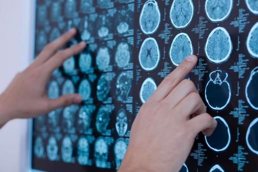
Magnetic resonance imaging (MRI) in the diagnosis of multiple sclerosis
This instrumental exam provides the doctor with images and is probably the most suitable exam for diagnosing multiple sclerosis.
MRI highlights the alterations of water content in the body, which correspond to the areas where the myelin has been replaced with plaques and which, in this case, are found above all in the bridge, in the corpus callosum and around the ventricles.
The use of a contrast agent highlights local BBB alterations which usually precede the signs of disease exacerbation; moreover, MRI with contrast medium also allows to highlight any lesions to the optic nerve, although it is not an examination capable of highlighting spinal cord lesions, for which the analysis of the cerebro-spinal fluid is usually used.
Analysis of the cerebro-spinal fluid
Cerebrospinal fluid is the fluid in which both the brain and spinal cord are immersed; it is made up of proteins, sugars and salts and has the function of nourishment and protection against any trauma, absorbing shocks.
Analysis of this fluid can provide invaluable information about the condition of the spinal cord and brain.
Samples of cerebrospinal fluid are taken with a lumbar puncture.
The patient is made to lie down in the fetal position and the doctor, using a syringe with a needle inserted directly into the back, is able to take a sample of liquid to be analysed.
After the sampling, however, the patient is forced to remain motionless for at least 12 hours.
The analysis of the cerebrospinal fluid goes to investigate the count of the cells present, the levels of proteins and glucose, the colour, the presence of antibodies and their quantity and of a possible infectious agent.
XP test (Summoned Potentials)
This test is done to measure the transmission and interpretation time of sensory messages that travel through the nerves.
The existence of slowdowns in transmission can be a sign of multiple sclerosis, due to the demyelination process.
It can be considered the exam par excellence for diagnosing multiple sclerosis as it gives detailed information on the functionality of the nerves, which is not possible to obtain with other diagnostic means; furthermore, it is also able to take into consideration areas of the body that cannot be investigated with other diagnostic means, such as the optic nerves and the spinal cord.
Read Also
Emergency Live Even More…Live: Download The New Free App Of Your Newspaper For IOS And Android

Multiple Sclerosis: What Are The Symptoms Of MS?
Rehabilitation Therapies In The Treatment Of Systemic Sclerosis
ALS Could Be Stopped, Thanks To The #Icebucketchallenge
Relapsing-Remitting Multiple Sclerosis (RRMS) In Children, EU Approves Teriflunomide
ALS: New Genes Responsible For Amyotrophic Lateral Sclerosis Identified
What Is “Locked-In Syndrome” (LiS)?
Amyotrophic Lateral Sclerosis (ALS): Symptoms To Recognise The Disease
Multiple Sclerosis, What It Is, Symptoms, Diagnosis And Treatment
CT (Computed Axial Tomography): What It Is Used For
Positron Emission Tomography (PET): What It Is, How It Works And What It Is Used For
CT, MRI And PET Scans: What Are They For?
MRI, Magnetic Resonance Imaging Of The Heart: What Is It And Why Is It Important?
Urethrocistoscopy: What It Is And How Transurethral Cystoscopy Is Performed
What Is Echocolordoppler Of The Supra-Aortic Trunks (Carotids)?
Surgery: Neuronavigation And Monitoring Of Brain Function
Robotic Surgery: Benefits And Risks
Refractive Surgery: What Is It For, How Is It Performed And What To Do?
Single Photon Emission Computed Tomography (SPECT): What It Is And When To Perform It