
Diagnostics: definition and function of fibroscopy
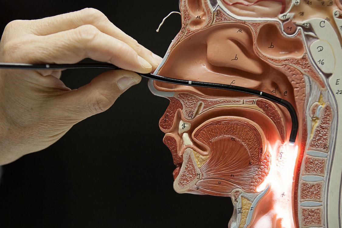
Fibroscopy is a diagnostic test that takes its name from the instrument with which it is performed, the fiberscope, i.e. an endoscope with the appearance of a thin tube, formed by very flexible optical fibers and equipped with a video camera connected to a monitor
What is Fibroscopy?
During the examination, the fiberscope is inserted into the interested cavity, returning, through the display of images transmitted on video, a detailed photograph of the area capable of identifying any anomalies.
This type of examination can take on a different name depending on the cavity involved.
In the case of an otorhinolaryngological examination, a distinction can be made between:
- fibrolaryngoscopy or laryngeal fibroscopy
- fiber rhinoscopy or nasal fibroscopy
- fiber endoscopy of the upper aerodigestive tract (when all structures from the nose to the hypopharynx are examined)
Laryngoscopy using a fiberscope allows you to analyze the pharynx, larynx and vocal cords, to evaluate the functionality of the organs, the motility of the vocal cords, the presence or absence of ulcerations, masses and inflammation in these areas.
This type of endoscopic examination is performed by inserting the optical fiber through the nasal cavity: once in position, the latter can examine the pharynx and larynx in detail.
It is good practice to practice it on an empty stomach or at least two hours after the last meal, but this is not an absolute recommendation.
This performance does not require anesthesia and is not painful.
Rhinoscopy, i.e. the inspection of the nasal cavities, takes place through the introduction of the fiberscope into the nose.
To avoid nausea, it is a survey that must be carried out on an empty stomach or at least two hours after the last meal.
Like laryngeal fibroscopy, nasal fibroscopy is painless and does not require anesthesia.
How is fibroscopy performed?
In general, no specific preparation is required from the patient to undergo a fibroscopy.
In the event that the subject has a particularly strong gag reflex (nausea), the otolaryngologist will apply a topical anesthetic locally in the form of a spray.
Although almost completely painless, this exam involves a slight discomfort, generated by the slight rubbing that the fiber produces against the nasal and/or pharyngeal mucosa.
In most cases the examination is well tolerated and for this reason it can be performed without the use of anesthesia.
During fibroscopy the patient will simply be asked to relax and breathe normally to allow a better view of the upper airways.
There are no contraindications to performing the examination in pediatric children.
The procedure is similar to that used for adults, the only difference lies in the size of the fiberscope which is smaller.

For the child, typically, fibroscopy is essential in the diagnosis of nasal obstructive pathologies such as adenoid hypertrophy.
What is it for
Specifically, fibroscopy is used in subjects with throat, nose, pharynx and larynx disorders.
It is common to resort to fibroscopy for the diagnosis of:
- sinusitis, a disease characterized by inflammation of the sinuses, cavities located behind the forehead, cheeks and eyes. It arises as a result of an infection with viruses, fungi or bacteria. It can be of two main types, acute when symptoms persist for up to a month, or chronic when it lasts for more than three months.
- hypertrophies of the turbinates, i.e. of the three bony folds located inside each nasal cavity (equipped with an anterior end called the head, and a posterior one, called the tail). The turbinates play an essential role in carrying out the functions of conditioning, heating, filtering and humidifying the inspired air. They are covered by a largely vascularized respiratory type mucosa, capable of modifying its volume in response to external stimuli of various kinds (irritant, phlogistic agents, traumas). However, when this increase in size stabilizes over time by more or less completely obstructing the nasal cavities, the full-blown pathological picture of hypertrophy is established, which most commonly involves the inferior turbinates and causes the typical sensation of a “stuffy nose”.
- sleep apnea, which is the interruption of a person’s regular breathing while sleeping. When the phenomenon occurs frequently, a syndrome called Sleep Apnea Syndrome (OSAS, Obstructive Sleep Apnea Syndrome) develops, which is a pathology characterized by a set of symptoms and disorders that are caused by recurrent breathing interruptions during sleep. The most characteristic symptoms of this pathology are: snoring, daytime tiredness, urinary incontinence, poor concentration ability, unstable mood, sudden falling asleep, dry mouth upon awakening, dysmnesia (i.e. memory disorders)
- tumors, in particular video-fibrolaryngoscopy with “Narrow-Band Imaging” (acronym NBI, narrow-band imaging) represents a very useful tool in the early diagnosis of precancerous lesions or early stage tumors affecting the larynx
- nasal polyps, i.e. semi-transparent formations that can form in any part of the naso-sinus mucosa following an inflammatory process of the mucosa. They are benign but can also block the nasal passages if they increase in size. They can develop in different degrees. At an early stage there may be a loss of smell and taste that can progress to partial or complete nasal obstruction. To this can be added a constant sensation of mucus which is relieved only by blowing the nose. When the polyps are numerous or large, rhinorrhea, snoring, a feeling of pressure in the forehead or even a headache may occur
- deviation of the nasal septum
- dysphonia, i.e. a disorder characterized by an alteration of the timbre of the voice, which can be attributable to various organic or functional causes of the larynx and vocal cords. Chronic aphonia in children and adults requires a doctor’s visit to diagnose any disturbance lasting more than 2 weeks, both to rule out serious injury and to prevent the hoarseness from turning into chronic aphonia and causing total voice loss
- vocal cord nodules, a condition where small bumps or swellings form on the edge of the vocal cords. The appearance of these nodules affects the glottic closure of the strings and is manifested by an alteration of the timbre of the voice, which is broken and more intense. Vocal cord nodules are generally not problematic, as they respond well to treatment, whether it is voice rehabilitation or surgery
- cyst
- gastroesophageal reflux, i.e. the ascent of gastric juices from the stomach into the esophagus, the channel through which liquids and foods flow from the mouth to the stomach. Gastroesophageal reflux disease (GERD) occurs when symptoms occur several times during the day and are associated with other ailments.
Other less common applications consist in the exploration of the glottis and trachea, as well as the bronchi in patients who have a tracheostomy tube.
Read Also
Emergency Live Even More…Live: Download The New Free App Of Your Newspaper For IOS And Android
Esophageal Mobility: What Is The FLIP Exam?
Flip, The New Test For Oesophageal Motility Disorders
Esophagogastroduodenoscopy (EGD Test): How It Is Performed
Gastro-Oesophageal Reflux Disease (GERD): Symptoms, Diagnosis And Treatment
What Is Esophagogastroduodenoscopy?
Indigestion Or Dyspepsia, What To Do? The New Guidelines
Dyspepsia: What It Is, Symptoms, Diagnosis And Treatment
Gastro-Oesophageal Reflux: Symptoms, Diagnosis And Treatment
Functional Dyspepsia: Symptoms, Tests And Treatment
Straight Leg Raise: The New Manoeuvre To Diagnose Gastro-Oesophageal Reflux Disease
Gastroenterology: Endoscopic Treatment For Gastro-Oesophageal Reflux
Oesophagitis: Symptoms, Diagnosis And Treatment