
Eyelid ptosis: an overview of the drooping eyelid
While the term ‘ptosis’ generally indicates the displacement of a physical structure due to the force of gravity, and can affect different parts of the body, eyelid ptosis is the most common
Those who suffer from it speak of ‘drooping eyelid’, as the eye seems to close: the pupil is obscured, sometimes only partially, sometimes totally, and the only way to solve the problem (not only aesthetically) is surgery.
Typical of ageing, eyelid ptosis can also affect children.
And it can have numerous causes.
What is eyelid ptosis?
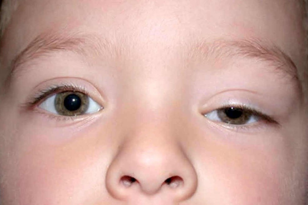
Eyelid ptosis is the partial or total drooping of the lower or upper eyelid.
It may be unilateral and thus affect only one eye, or bilateral and affect both.
Ptosis is mild if the drooping is less than 2 millimetres, moderate if it is between 2 and 4 millimetres, and severe if it is more than 4 millimetres.
It can also be congenital if it is present from birth or acquired if it appears later.
If in children it is caused by dystrophy of the muscle responsible for lifting the eyelid, or a neurological deficit, in adults and the elderly the cause is usually senile involution of the tendon of the muscle that lifts the eyelid.
As a rule, eyelid ptosis does not conceal other pathologies or is due to trauma.
In rare cases, however, it can be caused by muscular or neurological diseases or tumours.
Causes
In order to prevent the eyelid from ‘falling down’, all the structures that hold it in place must work perfectly: the upper eyelid elevator muscle, the orbicularis muscle, the neuromuscular plate and the Müller muscle (upper tarsal muscle).
When each of them does its job, the upper eyelid margin stops 1-2 millimetres above the cornea and has a distance of 9-10 millimetres to the lower eyelid.
Otherwise, eyelid ptosis occurs.
The main distinction is made between congenital and acquired ptosis, based on its causes.
Congenital ptosis
Congenital ptosis is a condition that is present from birth, and is usually caused by incomplete development of the elevator muscle.
Sometimes, it can be caused by a genetic or chromosomal defect or a neurological dysfunction.
There are several subcategories:
- Simple congenital ptosis is the most frequent and can manifest itself with varying degrees of intensity. In order to compensate for the incomplete development of the elevator muscle, the child contracts the frontalis muscle and tends to move the head to the side, risking compromising the curvature of the spinal column or generating strabismus (which is why rapid intervention is necessary to correct the ptosis);
- We speak of congenital ptosis related to abnormalities of oculo-palpebral motility when the problem is due to insufficient activity of the superior rectus muscle, congenital paralysis of the third cranial nerve, Marcus Gunn syndrome (sufferers involuntarily retract the eyelid when opening their mouth) or a malformation.
Acquired ptosis
Acquired ptosis occurs during adult life, and is in most cases due to a normal ageing process.
Neurogenic ptosis can have central or peripheral origins.
In the former case, it is often due to lesions of the frontal or temporal lobe, and is accompanied by paralysis of the muscles contained within the orbital cavity; in the latter case, it is caused by an impairment of the third cranial nerve.
Myogenic ptosis may be senile or, more rarely, related to myopathic syndromes.
The former are caused by an involution of the muscle fibres of the elevator muscle and Muller’s muscle (upper tarsal muscle, involved in eyelid movement), the latter are much less frequent and are due to rare pathologies (Steinert’s disease, Basedow’s disease, etc.).
Aponeuretic ptosis generally occurs in predisposed subjects due to trauma or following surgery (for retinal detachment, cataracts), and is due to the opening or disconnection of the aponeurosis (the tendon of the muscle that elevates the eyelid).
Mechanical ptosis is caused by formations on the eyelid due to benign or malignant tumours, scarring or oedema.
Traumatic ptosis is caused, as the name suggests, by blunt trauma or a lacerated wound.
Neurotoxic ptosis is due to poisoning and, since it is often accompanied by other serious symptoms, must be treated as an emergency.
Among the diseases that most frequently cause eyelid ptosis are
- myasthenia gravis, a condition that causes severe muscle weakness;
- foetal alcohol syndrome, a serious fetal condition caused by alcohol consumed by the mother during pregnancy;
- congenital anomalies;
- infections or inflammations of the eyelid;
- mental retardation;
- muscular dystrophies;
- tumours;
- strokes;
- diabetes;
Symptoms
Eyelid ptosis is itself a symptom.
The patient realises that he or she is suffering from it because the upper eyelid, of one or both eyes, falls to cover the eye.
It may be a slow process, or it may appear suddenly, and may be barely noticeable or cover the pupil entirely, obstructing or preventing vision.
Sometimes, the person may experience other symptoms such as difficulty opening and closing the eye, a sagging of the skin over the eyelid and pain around the eyes.
If a child suffers from ptosis, he or she usually raises the eyebrows or lifts the head backwards to try to see better, and may experience headaches or a stiff neck.
The most serious consequence of eyelid ptosis is amblyopia (or ‘lazy eye’), a more or less severe reduction in visual capacity.
The diagnosis of eyelid ptosis is made by the ophthalmologist
The examination consists of a palpation of the eyelid and palpebral orbit (the cavity that contains the eye, protecting it).
Afterwards, the specialist will proceed with measuring the distance between the upper and lower eyelid, and between the centre of the pupillary reflex to light and the lower and upper eyelid margin; he will also assess the functional capacity of the elevator muscle and the distance from the upper eyelid margin to the skin fold.
The ophthalmologist’s task is to assess the situation in the round, making sure that the patient performs eye movements correctly, produces adequate tearing and that the eyelid rhyme closes correctly.
He/she will also have to rule out the presence of other pathologies such as thyroid orbitopathy (a disease related to a malfunctioning thyroid gland), dermatocalasis (excess skin on the eyelid, which occurs when the connective tissue loses elasticity), entropion (the eyelid margin is turned inwards and irritates the cornea) or ectropion (the eyelid margin is turned outwards, causing irritation of the conjunctiva).
Once eyelid ptosis has been diagnosed, he will determine its severity and prescribe further investigations to investigate its causes.
He will then check for a neurological disorder, the possible presence of a mass within the eye cavity, and possibly request an imaging test (MRI or CT scan).
Treatment of ptosis depends on its severity and causes
If the ptosis is congenital and mild, without amblyopia or problems such as strabismus or head curvature, periodic monitoring is usually sufficient.
If deemed appropriate, the specialist may prescribe specific eye exercises to strengthen the muscles, eyeglasses for eyelid ptosis or contact lenses for eyelid support.
More severe cases of eyelid ptosis require surgery.
The mode of intervention is decided on the basis of the severity of the ptosis and its cause:

- if the elevator muscle needs to be strengthened, its tendon will be shortened or reinserted;
- if the elevator muscle cannot be strengthened, autologous or heterologous material is used to suspend the eyelid from the frontalis muscle;
- to reinforce Muller’s muscle, or to advance the aponeurosis, the transconjunctival technique is applicable without external incisions, but only in cases of mild eyelid ptosis.
With a dual effect, both aesthetic and functional, the surgery is followed by the application of some ice or a slightly compressive bandage.
For the first 24 hours, the patient must keep his head elevated. And, for about ten to twenty days, the skin may be reddened, swollen and bruised.
Vision may be blurred or double, and there may be a tendency to tearing and increased sensitivity to light.
Small haemorrhages may occur under the conjunctiva, but these tend to reabsorb spontaneously after a few days.
Possible complications of surgical correction of eyelid ptosis are
- infections requiring antibiotic therapy
- excessive eyelid retraction, which can usually be resolved with a specific massage but sometimes requires a further operation;
- lagophthalmos (the patient cannot close the eye properly and, if artificial tears do not solve the problem, needs further surgery);
- loss of eyelid sensitivity, which usually resolves spontaneously within three months;
- dryness of the eye, which makes the use of lubricating eye drops necessary;
- raised scars;
- wound opening and bleeding;
- formation of haematomas that must be surgically drained.
In any case, it is a good idea, after the operation, to avoid driving for a few days, exertion for the first few weeks, wearing contact lenses for at least fifteen days and sunbathing for two months.
The surgeon will assess when to remove the stitches, and will prescribe the therapy to follow based on ointments and pain-relieving eye drops, antibiotics and lubricants.
Surgery, however, is reserved for more serious cases of eyelid ptosis, in which the patient has a reduced field of vision, has assumed a spoiled head and neck posture, often has headaches due to the habit of frowning in order to see better, and looks tired.
In other cases, a non-surgical modality of intervention tends to be preferred.
Read Also
Emergency Live Even More…Live: Download The New Free App Of Your Newspaper For IOS And Android
Causes, Remedies And Exercises For Eyelid Ptosis
Blepharoptosis: Getting To Know Eyelid Drooping
Pupillary Reflex To Light: Mechanism And Clinical Significance
4 Reasons To Seek Emergency Care For Vision Symptoms
Eye Diseases: What Is Iridocyclitis?
Conjunctival Hyperemia: What Is It?
Eye Diseases: The Macular Hole
What Is Ocular Pterygium And When Surgery Is Necessary
Tear Film Dysfunction Syndrome, The Other Name For Dry Eye Syndrome
Vitreous Detachment: What It Is, What Consequences It Has
Macular Degeneration: What It Is, Symptoms, Causes, Treatment
Conjunctivitis: What It Is, Symptoms And Treatment
How To Cure Allergic Conjunctivitis And Reduce Clinical Signs: The Tacrolimus Study
Bacterial Conjunctivitis: How To Manage This Very Contagious Disease
Allergic Conjunctivitis: An Overview Of This Eye Infection
Keratoconjunctivitis: Symptoms, Diagnosis And Treatment Of This Inflammation Of The Eye
Glaucoma: What Is True And What Is False?
Eye Health: Prevent Conjunctivitis, Blepharitis, Chalazions And Allergies With Eye Wipes
What Is Ocular Tonometry And When Should It Be Done?
Dry Eye Syndrome: How To Protect Your Eyes From PC Exposure
Autoimmune Diseases: The Sand In The Eyes Of Sjögren’s Syndrome
Dry Eye Syndrome: Symptoms, Causes And Remedies
How To Prevent Dry Eyes During Winter: Tips
Blepharitis: The Inflammation Of The Eyelids
Blepharitis: What Is It And What Are The Most Common Symptoms?
Stye, An Eye Inflammation That Affects Young And Old Alike
Diplopia: Forms, Causes And Treatment
Exophthalmos: Definition, Symptoms, Causes And Treatment
Eye Diseases, What Is Entropion
Hemianopsia: What It Is, Disease, Symptoms, Treatment
Diseases Of The Ocular Conjunctiva: What Are Pinguecula And Pterygium And How To Treat Them
Ocular Herpes: Definition, Causes, Symptoms, Diagnosis And Treatment
Eye Diseases: What Is Iridocyclitis?
Hypermetropia: What Is It And How Can This Visual Defect Be Corrected?
Miosis: Definition, Symptoms, Diagnosis And Treatment
Floaters, The Vision Of Floating Bodies (Or Flying Flies)
Nystagmus: Definition, Causes, Symptoms, Diagnosis And Treatment