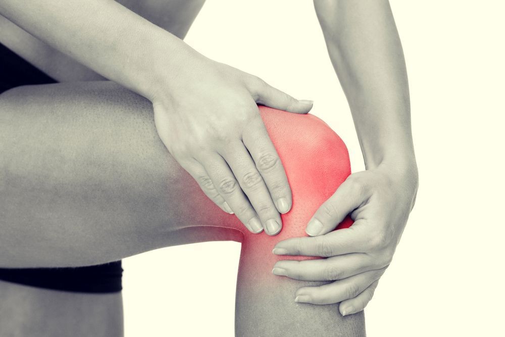
Knee pathologies: the synovial plica
The synovial plica is a thickening of the synovial membrane of the knee, a thin connective tissue membrane present in the joints
Under normal conditions, there are four main folds in the knee, but the one most frequently responsible for a disorder is the medial patellar fold.
When we speak of thickening of the synovial flap, we are referring to a frequent but often asymptomatic disorder that occurs when the flap, as it thickens, becomes incarcerated between hard structures.
When the flap then becomes inflamed, pain may occur in some cases, causing difficulty in sliding the bony joints.
Data have shown that the female sex is more affected than the male by this disorder.
Synovial fold thickening can be idiopathic, or secondary to trauma or inflammation of the synovial tissue.
Secondary forms are the most frequent and are often due to repeated trauma or microtrauma during sporting activity and repetitive flexion-extension movements, as is often the case in those doing activities such as running.
The diagnosis of synovial plica is made through clinical evaluation by the doctor and by performing various tests, but it is only certain after arthroscopic evaluation
Sometimes it is enough to modify the physical activity performed, e.g. replace jogging with cycling, and if there is a particularly sensitive area of the knee, apply ice after physical activity.
In uncomplicated cases, a slow recovery can be achieved within 3-6 months.
The treatment is initially conservative, i.e. it involves medical therapy and physiotherapy for a few weeks, and when the latter fails, the plica is removed arthroscopically with special scissors and a shaver.
It should be remembered that surgery is reserved for those patients in whom all conservative treatment modalities have failed.
No particular complications have been reported, although it remains very important to diagnose this condition in order to eliminate any discomfort perceived by those affected.
Synovial plica symptoms
Synovial flap symptoms are mainly knee pain during flexion-extension movements of the joint and episodes of pseudo-joint locking due to the presence of inflammation and joint effusion.
In reality, synovial plica pain rarely occurs in isolation; it is usually associated with other conditions responsible for knee pain syndromes, such as a meniscus rupture, the outcomes of Osgood Schlatter’s disease (inflammation of the tibial tuberosity among the most common causes of knee pain) and patellar tendinitis.
A person with synovial plica may also feel a sensation of tension along the inside of the knee when it is bent.
In this position, in fact, one can feel pain when touching and the plica can also be felt under the skin as a string that snaps during knee movement.
In a few cases the knee, especially if the plica is very inflamed, may also appear swollen due to the inflammatory effusion inside it.
Sometimes the subject perceives a ‘morning pop’ present upon waking during flexion-extension of the knee at about 30° to 40° of flexion and which then disappears during the day.
Causes of synovial plica
In practice, repetitive flexion and extension movements of the knee, such as in running, but also in typical everyday postures such as climbing and descending stairs, stepping or squatting can cause this inflammation.
From an anatomical point of view, there are four types of knee folds
- suprapatellar plica
- mediopatellar fold
- parapatellar fold
- infrapatellar plica
The plica that most frequently causes inflammatory symptoms is the mediopatellar plica.
Chronic inflammation can cause a condition of fibrosis, which consequently causes the plica to lose elasticity and become inflexible and thicken.
The result is that the hardened plica is trapped between the patella and the femoral condyle during the extension movement, causing pain in the patient.
One of the rather rare causes of this condition could be due to hypertone, stiffness or hypertrophy of the genu articularis muscle whose action is to avoid trapping the capsule between the patella and trochlea while extending the knee.
Excessive pulling by this muscle leads to increased tension of the plica, which increases rubbing friction on the condyle during knee flexion.
Diagnosis
The diagnosis of the synovial flap is basically clinical, i.e. based on the doctor’s test of the knee and the patient’s medical history.
Some authors have classified this lesion (synovial flap) into 4 types:
- TYPE A: chordiform flap
- TYPE B: narrow flap
- TYPE C: wide flap
- TYPE D: subdivided into three subgroups: fenestrated, double and broken bucket-handle plexus
To identify a synovial fold, several tests are performed, namely
- the extension test: with the patient lying supine and the knee flexed by 90°, a quick extension of the tibia is requested as if kicking a ball and the test is considered positive if this movement causes pain;
- the flexion test: with the patient supine, knee extended and off the couch, the patient is asked to do a quick flexion then an abrupt stop when the knee is flexed approximately 30°-60°, if this manoeuvre causes pain the test is considered positive;
- the MMP test: consists of the manual application of stress to the inferior medial patellofemoral joint with the patient supine with the examiner’s thumb, which identifies the presence of pain. If this pain decreases with 90° flexion while maintaining force, the MPP test is considered positive.
Other tests that may be required for diagnosis are the rotation valgus test and the Koshino and Okamoto holding test, however they have a lower level of reliability. From an instrumental point of view, an MRI can provide the physician with a lot of valuable information, but it may not be sufficient to distinguish a normal from a pathological plica.
Conventional X-rays are necessary to eliminate other causes of knee pain, but they do not help in diagnosing plica.
In fact, X-rays are not necessary in most cases, partly because the flap is invisible to X-rays, but they may be required to check for other changes in the knee.
More help is provided by musculoskeletal ultrasound, which allows good visualisation of the soft tissues.
Pathologies to be considered in differential diagnosis include patellofemoral syndrome (most common) and medial meniscal injuries, but also lateral facet hyperpressure syndrome and Hoffa’s syndrome.
The definitive diagnosis requires a rather invasive test, i.e. arthroscopy, which consists of inserting a fibre-optic cable connected to a camera and a television monitor inside the knee, allowing the inside of the knee to be seen.
Apart from recurrences, complications in arthroscopic surgery of the plica syndrome are absent or few.
They are listed below:
- superficial infections
- septic arthritis
- joint effusions
- deep vein thrombosis
- pulmonary embolism
- iatrogenic nerve damage
- damage due to iatrogenic lesions of vesselskeloid scars
- failure due to pain on meniscal residuum
- persistent pain
- iatrogenic cartilage injuries
- knee stiffness
- lameness
- hemarthrosis
Treatments
Therapy for synovial plica is aimed at regressing the acute or inflammatory phase, by resting the limb and taking non-steroidal anti-inflammatory drugs, and then proceeding with physiotherapy involving quadriceps strengthening and stretching exercises to manage muscle and joint recovery.
Usually the subject does not need to use crutches.
Physical therapy techniques (laser, ultrasound and iontophoresis, etc.) can also be used to eliminate the inflammation, and infiltrations with cortisone into the plica can be useful.
Generally, with these measures, the symptoms tend to recede. If, on the other hand, the symptoms are persistent, the doctor may recommend surgery, which is only required in certain cases, i.e:
- when symptoms are certainly consequent to the presence of the synovial flap, in the absence of other joint pathologies that could justify it;
- when a highly fibrotic plica is found, as conservative therapy is less effective in these cases;
- if there are chondral plication lesions (i.e. grooves on the femoral condyle caused by contact with the plication).
Complications of synovial plica surgery
Intervening on the synovial flap is important because in severe cases it can evolve into a fibrotic, cord-like flap.
In order to avoid the complications that can result from this condition, it is very important that those who are more exposed, such as runners, correct any excess pronation by adopting models of shoes with greater stability and possibly also anti-shock insoles or orthotics.
Read Also
Emergency Live Even More…Live: Download The New Free App Of Your Newspaper For IOS And Android
Rotator Cuff Injury: What Does It Mean?
Tendon Injuries: What They Are And Why They Occur
Elbow Dislocation: Evaluation Of Different Degrees, Patient Treatment And Prevention
Cruciate Ligament: Watch Out For Ski Injuries
Sport And Muscle Injury Calf Injury Symptomatology
Meniscus, How Do You Deal With Meniscal Injuries?
Meniscus Injury: Symptoms, Treatment And Recovery Time
First Aid: Treatment For ACL (Anterior Cruciate Ligament) Tears
Anterior Cruciate Ligament Injury: Symptoms, Diagnosis And Treatment
Work-Related Musculoskeletal Disorders: We Can All Be Affected
Patellar Luxation: Causes, Symptoms, Diagnosis And Treatment
Arthrosis Of The Knee: An Overview Of Gonarthrosis
Varus Knee: What Is It And How Is It Treated?
Patellar Chondropathy: Definition, Symptoms, Causes, Diagnosis And Treatment Of Jumper’s Knee
Jumping Knee: Symptoms, Diagnosis And Treatment Of Patellar Tendinopathy
Symptoms And Causes Of Patella Chondropathy
Unicompartmental Prosthesis: The Answer To Gonarthrosis
Anterior Cruciate Ligament Injury: Symptoms, Diagnosis And Treatment
Ligaments Injuries: Symptoms, Diagnosis And Treatment
Knee Arthrosis (Gonarthrosis): The Various Types Of ‘Customised’ Prosthesis
Rotator Cuff Injuries: New Minimally Invasive Therapies
Knee Ligament Rupture: Symptoms And Causes
MOP Hip Implant: What Is It And What Are The Advantages Of Metal On Polyethylene
Hip Pain: Causes, Symptoms, Diagnosis, Complications, And Treatment
Hip Osteoarthritis: What Is Coxarthrosis
Why It Comes And How To Relieve Hip Pain
Hip Arthritis In The Young: Cartilage Degeneration Of The Coxofemoral Joint
Visualizing Pain: Injuries From Whiplash Made Visible With New Scanning Approach
Coxalgia: What Is It And What Is The Surgery To Resolve Hip Pain?
Lumbago: What It Is And How To Treat It
Lumbar Puncture: What Is A LP?
General Or Local A.? Discover The Different Types
Intubation Under A.: How Does It Work?
How Does Loco-Regional Anaesthesia Work?
Are Anaesthesiologists Fundamental For Air Ambulance Medicine?
Epidural For Pain Relief After Surgery
Lumbar Puncture: What Is A Spinal Tap?
Lumbar Puncture (Spinal Tap): What It Consists Of, What It Is Used For
What Is Lumbar Stenosis And How To Treat It
Lumbar Spinal Stenosis: Definition, Causes, Symptoms, Diagnosis And Treatment
Cruciate Ligament Injury Or Rupture: An Overview
Haglund’s Disease: Causes, Symptoms, Diagnosis And Treatment
Osteochondrosis: Definition, Causes, Symptoms, Diagnosis And Treatment
Osteoporosis: How To Recognise And Treat It
About Osteoporosis: What Is A Bone Mineral Density Test?
Osteoporosis, What Are The Suspicious Symptoms?
Osteoporosis: Definition, Symptoms, Diagnosis And Treatment
Back Pain: Is It Really A Medical Emergency?
Osteogenesis Imperfecta: Definition, Symptoms, Nursing And Medical Treatment
Exercise Addiction: Causes, Symptoms, Diagnosis And Treatment
Osteoarthrosis: Definition, Causes, Symptoms, Diagnosis And Treatment


