
Prenatal pathologies, congenital heart defects: Pulmonary atresia
Pulmonary atresia is an obstruction that prevents blood from being oxygenated in the lungs. It must be diagnosed before birth and corrected with a series of surgical interventions
Pulmonary atresia is a congenital heart defect of the pulmonary valve (present at birth)
The valve serves to allow blood directed to the lungs to flow out of the right ventricle.
In this disease, the pulmonary valve can be completely closed (complete atresia) or almost completely closed (critical pulmonary valve stenosis).
There are two forms of pulmonary atresia
They constitute two completely different diseases that also require different treatment modalities:
- Pulmonary atresia with an intact interventricular septum;
- Pulmonary atresia with interventricular septal defect.
In intrauterine life, according to the data available to us, this anomaly generally does not impair the child’s development because it is the placenta that supplies the body with the necessary oxygen instead of the lungs.
The blood entering the right side of the heart through a hole (foramen ovale) passes from the right atrium to the left atrium, then to the left ventricle and from there it can be pumped to the rest of the body through the aorta.
However, as it cannot reach the lungs via the normal route (right ventricle, then valve and pulmonary artery, then lungs) the blood reaches them via the ductus arteriosus, a vessel that connects the aorta to the pulmonary artery.
This structure is necessary during development in the womb and normally closes immediately after birth.
Therefore, if in the first few minutes of life the baby does not receive a drug that keeps the ductus arteriosus pervious – open – (prostaglandins, intravenously), the failure to pass blood from the lungs can be fatal.
This is why pulmonary atresia is a neonatal emergency
It is therefore essential that the diagnosis be made early: prenatal diagnosis allows the safest possible preparation for delivery and thus avoids dangerous delays in administering the necessary treatment.
Pulmonary atresia with intact ventricular septum
When there is no interventricular defect (DIV) – a formation defect in the wall, or septum, separating the two ventricles – the right ventricle receives little blood flow even before birth and its development may be impaired.
Under certain conditions (absence of tricuspid valve insufficiency), the right ventricle is usually very small and after birth is not fit to perform its function, i.e. to pump blood into the lungs.
This form is the most severe.
In other cases, however, e.g. when major tricuspid valve insufficiency is present, the ventricle may be well developed or bordeline (reduced in volume but not completely).
Pulmonary atresia with interventricular septal defect
When the interventricular defect is present, which allows the exchange of blood between the two ventricles, the right ventricle is usually well developed.
In this form, however, varying degrees of hypoplasia (reduced development) of the pulmonary arteries can occur until they are very small or non-existent.
In the latter case, additional sources of pulmonary flow called systemic-pulmonary collaterals or MAPCAs (acronym for major aorto-pulmonary collateral arteries) are formed. These vessels carry blood from the arterial circulation to the lungs and may be numerous, have a tortuous and variable course in the chest.
This form is the only one that does not represent a neonatal emergency.
Symptoms will be evident soon after birth
These may include:
- Cyanosis (bluish or greyish discolouration of the skin, caused by low oxygen content in the blood);
- Rapid breathing or shortness of breath;
- Difficulty in taking meals.
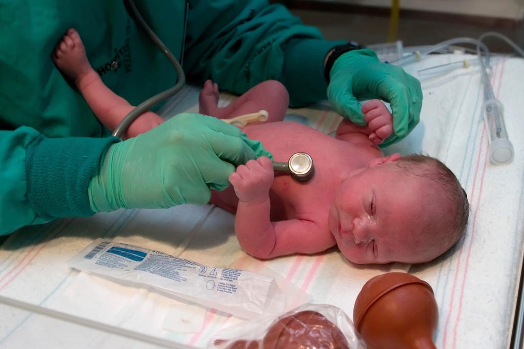
When the doctor suspects pulmonary atresia, the infant is sent to a paediatric cardiology centre.
The cardiologist has several means at his disposal to confirm the diagnosis.
Among these, the echocardiogram is the one that allows him to confirm the diagnosis.
The electrocardiogram (ECG) and chest X-ray can add important data.
Pulse oximetry indicates the amount of oxygen in the baby’s blood. In some cases, other examinations such as contrast-enhanced computed tomography or cardiac catheterisation are essential to define the anatomy of the pulmonary arteries and any additional sources of pulmonary flow (MAPCAs).
Today, it is possible to diagnose pulmonary atresia even during pregnancy thanks to the fetal echocardiogram
When the gynaecologist suspects this heart disease, he or she sends the pregnant woman to a specialised paediatric cardiology centre where the paediatric cardiologist confirms the diagnosis, explains to the parents the significance of the disease and the implications for life after birth, along with the treatment options.
The unborn child must then be born in a level II-III centre and transferred at birth to a paediatric cardiology and cardiac surgery centre for appropriate treatment.
Fetal diagnosis minimises the delay in starting appropriate treatment and improves the survival of these young patients.
Treatment of pulmonary atresia is necessarily surgical or interventional
Very often, in the first years of life, more than one surgical intervention is required.
Some are performed in the first days or weeks of the child’s life and others later.
In both forms of pulmonary atresia (with or without interventricular septal defect), the administration of intravenous prostaglandins at birth is essential for the child’s survival (except in the form with MAPCAs).
At this point, treatment changes depending on the disease variant.
How intact septal lung atresia is treated

To date, there are several strategies for the initial treatment of this disease.
The use of one or the other depends on the specific variant and the operators’ experience.
If the right ventricle is well formed, or it is believed that it will be able to grow adequately in the future and fully perform its pump function, procedures can be performed, either singly or in a variable combination, that maintain the functioning of the heart with two separate ventricles (biventricular circulation):
- Valve opening (valvulotomy) with a balloon or removal with radiofrequency: this is performed by cardiac catheterisation, a procedure in which catheters are inserted through the veins or arteries of the body to reach the heart. The aim is to create a small hole in the centre of the atresic valve and then open its leaflets with a balloon;
- Placement of stent in the ductus arteriosus: the stent is a small wire mesh tube that keeps the ductus arteriosus open. Stent placement is also performed via cardiac catheterisation and can be combined with valve opening (previous point);
- Systemic-pulmonary shunt and widening of the right ventricular outflow: this is a surgical procedure that works by creating a connection between one of the arteries arising from the aorta and the pulmonary artery (shunt) using a small tube of synthetic material (the most common example is called a Blalock-Taussig shunt orBT shunt), opening the pulmonary valve and widening the surrounding area. This procedure involves removing or closing the shunt after a few years (by cardiac catheterisation) and placing a new pulmonary valve either surgically or percutaneously in young adulthood.
If, on the other hand, the right ventricle is so small that it cannot perform its pumping function, it is necessary to operate the heart with only the left ventricle pumping oxygenated blood into the aorta; blood arriving from veins throughout the body will reach the lungs without passing through the right ventricle (monoventricular circulation).
This is a temporary remedy that requires at least three surgeries in the first few years of life (stage III):
- The first, in the first days of life, consists of the creation of the systemic-pulmonary shunt (stage I, see above);
- The second procedure, Glenn’s bidirectional hollow-pulmonary anastomosis, in which the surgeon attaches the blood vessel that collects blood from the upper part of the body (superior vena cava) to the pulmonary artery. This procedure is generally performed between 4 and 6 months of age (stage II) and the oxygen saturation in the blood remains below normal, around 85%;
- In the third, Fontan surgery, which is usually performed when the child is 2 or 3 years old, the surgeon will connect the blood vessel that collects blood from the lower part of the body (inferior vena cava) to the pulmonary artery. This serves to restore the oxygen level to 99% (stage III).
In the case of the borderline right ventricle, a one-and-a-half ventricle repair can be performed, which involves Glenn’s bidirectional cable-pulmonary anastomosis (connection between two organs) (see above).
This may be the final procedure if the right ventricle in the child’s heart is large enough and does not require the Fontan procedure, thus reducing the likelihood of long-term problems that may occur in this case.
It is worth mentioning pulmonary valve angioplasty in intrauterine life
Only in a few centres in the world and only in carefully selected cases is it possible to perform this procedure, which aims to improve the growth of the right ventricle.
The risks are not negligible and the probability of success depends on several factors that must be examined on a case-by-case basis.
It must be remembered that this intervention does not replace the above-mentioned procedures, which may still be necessary after birth depending on the variant of the disease.
How pulmonary atresia with interventricular septal defect is treated
There are different treatment paths depending on the anatomy of the pulmonary arteries and the presence or absence of MAPCAS.
If the pulmonary arteries are well developed, placement of a stent in the ductus arteriosus via cardiac catheterisation or surgical placement of a systemic-pulmonary shunt in the first few days of life is possible.
Subsequently, at around 3-6 months of age, corrective surgery can be performed, which consists of closing the ventricular septal defect and, depending on the anatomy of the right ventricular outflow, placing a prosthetic valve conduit between the right ventricle and the pulmonary artery or opening the pulmonary valve and enlarging the surrounding area (trans-annular patch).
If, on the other hand, the pulmonary arteries are hypoplastic or absent and MAPCAs are present, the surgeon will have to connect all of these to create the new pulmonary arteries (unifocalisation), close the interventricular defect and place a conduit between the pulmonary arteries and the right ventricle.
The timing and order in which these three steps are performed vary from case to case and may include:
- The temporary placement of a shunt;
- Several interventions staggered over time, usually from 6 months up to the first few years of life;
- The use of intermediate diagnostic and/or interventional investigations such as computed tomography or cardiac catheterisations between interventions.
Read Also:
Emergency Live Even More…Live: Download The New Free App Of Your Newspaper For IOS And Android
Congenital Pulmonary Valve Stenosis And Ebstein’s Anomaly
Scimitar Syndrome: Causes, Symptoms, Diagnosis, Treatment, Prognosis And Mortality
Carotid Stenosis: What Is It And What Are The Symptoms?
Cervical Stenosis: Symptoms, Causes, Diagnosis And Treatment
Coronary Angioplasty, What To Do Post-Operatively?
Heart Patients And Heat: Cardiologist’s Advice For A Safe Summer
US EMS Rescuers To Be Assisted By Paediatricians Through Virtual Reality (VR)
Silent Heart Attack: What Is Silent Myocardial Infarction And What Does It Entail?
Mitral Valve Diseases, The Advantages Of Mitral Valve Repair Surgery
Coronary Angioplasty, How Is The Procedure Performed?
What It Is And How To Recognise Abdominal Diastasis
Chronic Pain And Psychotherapy: The ACT Model Is Most Effective
Hiatal Hernia: What It Is And How To Diagnose It
Percutaneous Discectomy For Herniated Discs
What Is That Swelling? Everything You Need To Know About Inguinal Hernia
Symptoms And Causes Of Umbilical Hernia Pain
Congenital Diaphragmatic Hernia (CHD): What It Is, How To Treat It