Rectosigmoidoscopy and colonoscopy: what they are and when they are performed
Rectosigmoidoscopy is a diagnostic technique by which one can look into the rectum and sigma (hence the term rectosigmoidoscopy) to see if there is any lesion causing the patient’s discomfort
What is colonoscopy
Colonoscopy is an instrumental technique that, in addition to exploring the rectum and sigma, also studies the remaining segments of the colon.
We speak of total colonoscopy (pancolonoscopy) when all sections of the large intestine are explored, from the anus to the ileo-cecal valve.
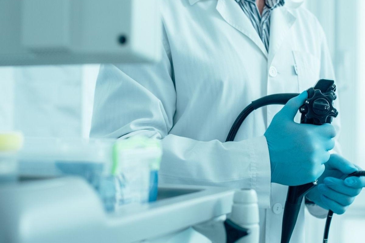
In both instrumental investigations, an endoscope is used, i.e. a flexible tube about a finger’s diameter with a bright light at its end that is passed through the anal canal into the colon.
Why and when rectosigmoidoscopy and colonoscopy are used
Rectosigmoidiscopy and colonoscopy are investigations that are performed when the patient has complaints or symptoms such as:
- persistent diarrhoea with or without emission of blood from the rectum (rectorrhagia)
- emission of mucus with the stool (mucorrhoea);
- belly pain;
- change in bowel habits;
- chronic anaemia without obvious pathology in the upper digestive tract.
In young patients (age ) with flu-like symptoms and/or with occasional episodes of rectal bleeding, at the doctor’s discretion, endoscopic exploration may also involve only the rectum and sigma if the presence of haemorrhoids is identified as the source of bleeding and if there are no other lesions in the explored tracts.
On the other hand, it becomes important to perform a total colonoscopy if inflammation is found in the rectum and sigma (e.g. ulcerative rectocholitis), if there is a polyp in the first tracts explored, if the subject is >40-45 years old and has rectal bleeding, if there is a family history of polyposis or bowel cancer.
However, a good gastroenterologist endoscopist, if the patient has a sufficiently clean bowel for adequate preparation, should always try to reach the ileo-cecal valve.
When it is not useful to do rectosigmoidoscopy and colonoscopy
Endoscopy certainly cannot resolve any functional or psychosomatic disorders for which the patient has been advised to have the examination.
In fact, the diagnosis of such disorders, labelled by the clinician as ‘functional symptoms’ or ‘irritable bowel symptoms’ (‘colitis nervosa’) is a diagnosis of exclusion (absence of pathology in the whole colon explored).
It is clear, however, that the absence of lesions on instrumental investigation often reduces the patient’s anxiety with alleviation or disappearance of his symptoms.
What do I need to know about rectosigmoidoscopy?
Preparation for rectosigmoidoscopy or colonoscopy is decisive for the success of the examination and it is therefore essential that it be performed correctly.
For a clear view, the colon must be completely free of faeces.
It is therefore necessary to take a laxative solution to drink the day before the investigation or in any case no less than 6 hours before the examination.
A light supper (soup, broth) can be taken the evening before.
Usually the endoscopic investigation of the large intestine is unpleasant and sometimes a little painful.
Sometimes the pain may not be tolerable (usually this is caused by the anatomical conformation of the intestine, or scars from previous surgery on the belly, or the presence of large inguinal hernias; in this case medication may be administered to better tolerate the examination and the associated procedures.
What are the risks of these instrumental examinations?
When used for diagnostic purposes, by specially trained and experienced physicians, instrumental investigation of the colon is safe and associated with very few risks.
On the other hand, these are increased in operative endoscopy such as in the removal of a polyp (polypectomy).
The other problem concerns the potential transmissibility of infections, in particular hepatitis B, C, D, and AIDS viruses.
The possibility of transmitting infections by means of the endoscopic instrument is intuitive: the instrument in fact comes into contact with mucous membranes and accessories and the integrity of the mucosal barrier may be breached, especially during operative manoeuvres.
This possibility is closely related to improper cleaning and disinfection.
In fact, until new evidence comes to light, although possible, transmission of these viruses in endoscopy is infrequent and remains linked to failure to observe and incomplete observation of instrument cleaning and disinfection standards.
In fact, the cleaning and disinfection guidelines have now been defined internationally, guarantee a standard of decontamination with elimination of viruses, bacteria, fungi and therefore an almost zero risk of contagion.
Before the examination, you must carry out the preparation that has been indicated to you so that your intestines are perfectly clean to allow the operator an optimal view.
If this is not the case, the examination may take longer, may not be diagnostic or may be incomplete, hence the risk of repeating the examination after more careful preparation.
It is also important to bring any previous radiological examinations or colonoscopy reports to the doctor before he/she performs the examination.
Each patient participates in the investigation with a different psycho-emotional make-up and therefore even the same examination elicits different reactions in them.
How it is performed
The patient is placed on a couch on the left side.
After exploration of the anal canal with the operator’s finger, the instrument is introduced into the rectal ampulla and continued as far as possible to the end of the large intestine.
The chances of success depend on the cleanliness, the conformation of the intestine and the cooperation of the patient.
Air will be injected in order to stretch the walls of the intestine and have a better view, and this may cause some discomfort.
In fact, one may feel the sensation of ‘draining’ or feel a ‘bloated belly’ or complain of pain with abdominal cramps.
It is important to inform the staff present of your complaints, who will act accordingly.
The examination can last from a few minutes (if only the rectum and sigma are explored) to 15-30 minutes if a total colonoscopy is performed.
Overall, the complication rate during diagnostic endoscopy is less than 4 per thousand.
It is clear that patients with concomitant diseases, such as cardiovascular, pulmonary, renal, severe hepatic, neurological and metabolic diseases, as well as advanced age, have a higher risk of complications.
During the examination it is possible to encounter intestinal polyps.
These are protuberances (outgrowths) of the mucosa of the intestinal wall facing the lumen that have a tendency to increase in volume (from a few mm to several cm) over time.
They can also give rise to certain complications such as bleeding, intestinal obstruction, but above all in some cases they can develop into malignant tumours.
It is therefore prudent, whenever a polyp is found during a colonoscopy, to remove it, have it analysed under a microscope (histological examination) and schedule periodic surveillance.
This is why it is necessary to remove polyps (polypectomy); this can be done during rectosigmoidoscopy or colonoscopy
All patients who present with polyps, who are not wearers of cardiac pacemakers and who have normal blood coagulation can undergo polypectomy.
In this regard, since polyps are relatively frequently observed during the endoscopic examination, it is advisable for patients over the age of 45 or patients known to have polyposis (personal or family) to have laboratory tests a few days before the investigation to assess their coagulation status (blood count, fibrinogen, platelets, prothrombin time, partial thromboplastin time).
In this way, if a polyp is observed during the endoscopic examination and there is a possibility of one, it will be removed immediately in order to prevent the patient from having to undergo another endoscopy.
Is polypectomy dangerous?
No, it is not a dangerous procedure; the removal of polyps is painless.
However, it must be considered that it is a real surgical procedure and as such carries risks.
In this regard, the patient will be asked to sign a sheet, the so-called ‘informed consent’, i.e. a statement in which he or she consents to the doctor performing the operative procedure.
This consent does not exempt the doctor from his professional responsibilities.
Complications are possible in about 1% of cases.
Such complications are:
- haemorrhage, which usually clears up on its own, but still requires hospitalisation for observation, although surgery is rarely necessary;
- perforation of the intestine, which always requires corrective surgery.
What the patient should do after the endoscopic examination
At the end of the investigation, after a few minutes’ rest, the patient should go home.
The report of the endoscopy will be given to him immediately, while he will have to wait 5 to 10 days for the results of any biopsies (histological examination).
In the case of a polypectomy, the patient remains under observation for 30 to 60 minutes and, at the doctor’s discretion, may possibly be invited for a short hospitalisation if a complication is suspected.
If he or she has been given sedation medication, it is important that a companion is available to drive him or her home, as sedation impairs reflexes and judgement.
For the remainder of the day, you will not be able to drive a car, operate machinery or make important decisions.
It is advisable to remain at rest for the entire day.
Sedation commonly refers to a drug-induced reduction in the level of consciousness to facilitate the acceptability of endoscopic investigation.
The most commonly used drugs are benzodiazepines that induce relaxation and cooperation on the part of the patient, and in some cases even a state of amnesia.
Read Also
Emergency Live Even More…Live: Download The New Free App Of Your Newspaper For IOS And Android
Bone Scintigraphy: How It Is Performed
Fusion Prostate Biopsy: How The Examination Is Performed
CT (Computed Axial Tomography): What It Is Used For
What Is An ECG And When To Do An Electrocardiogram
Positron Emission Tomography (PET): What It Is, How It Works And What It Is Used For
Single Photon Emission Computed Tomography (SPECT): What It Is And When To Perform It
Instrumental Examinations: What Is The Colour Doppler Echocardiogram?
Coronarography, What Is This Examination?
CT, MRI And PET Scans: What Are They For?
MRI, Magnetic Resonance Imaging Of The Heart: What Is It And Why Is It Important?
Urethrocistoscopy: What It Is And How Transurethral Cystoscopy Is Performed
What Is Echocolordoppler Of The Supra-Aortic Trunks (Carotids)?
Surgery: Neuronavigation And Monitoring Of Brain Function
Robotic Surgery: Benefits And Risks
Refractive Surgery: What Is It For, How Is It Performed And What To Do?


