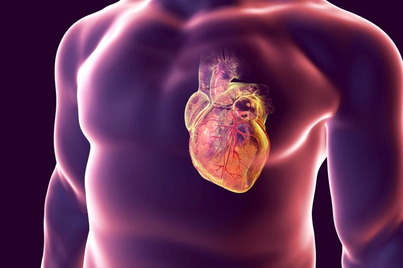
Cardiac arrest: what it is, what the symptoms are and how to intervene
Also known as ‘sudden cardiac death’, cardiac arrest is not so much a disease as a serious emergency situation that can lead to the immediate death of the individual
The heart of the person affected stops pumping blood, and therefore stops functioning: as a result of the absence of blood circulation, the person loses consciousness and stops breathing.
Only the timely and lucid intervention of medical personnel can save his life.
Although these are two very serious events, both affecting the heart, cardiac arrest and cardiac infarction should not be confused
In the first case, the patient loses consciousness within a few seconds, in the second he may remain conscious (the loss of consciousness during cardiac infarction depends on the extent of the damage generated by the ischaemia).
In both cases, it is absolutely necessary to seek immediate medical attention: in the case of a heart attack, the doctor’s intervention will be aimed at ‘opening’ the obstructed coronary artery, usually by means of an angioplasty or the administration of specific drugs; in the case of cardiac arrest, he will have to restart the heart by means of cardiopulmonary resuscitation.
More common with advancing age, cardiac arrest causes around 50,000 deaths a year in Italy (source: Ministry of Health). The survival rate is very limited: only 2% of people who do not undergo treatment survive, a rate that rises to 50% if the manoeuvres are correctly performed within five minutes of the onset of arrest.
What is cardiac arrest?
Cardiac arrest is an emergency situation, which arises suddenly and is characterised by the cessation of cardiac organ activity.
The blood stops circulating, the patient loses consciousness and reaches death in about an hour from the onset of symptoms if not treated promptly with specific resuscitation techniques.
The cause of cardiac arrest is almost always an arrhythmia, i.e. an alteration in cardiac rhythm.
The myocardium, the muscle of the heart that generates the impulses for the contraction of the atria and ventricles, is ‘managed’ by the atrial sinus node.
This is the source of the impulses, and its function is precisely to maintain a normal heart rhythm (or sinus rhythm) by regulating the rate of contraction.
When an arrhythmia is present, the heart beats faster or slower: in both cases, normal heart function is seriously impaired.
Cardiac arrest is therefore a sudden emergency, three times more frequent in men than in women and capable of leading to death within minutes.
The cause of the arrest is an arrhythmia, but it should be specified that not all arrhythmias lead to such an event
Arrhythmias are numerous and differ in terms of evolution and prognosis.
Bradyarrhythmias lead to a slower-than-normal heart rhythm, tachyarrhythmias to a faster rhythm.
Extrasystoles are characterised by an earlier beat, and are often of no clinical relevance; WPW syndrome is characterised by tachyarrhythmias due to the presence of an accessory bundle that sends the electrical impulse to abnormal areas of the heart.
Among the arrhythmias that most often lead to cardiac arrest is ventricular fibrillation, in the presence of which the ventricles fail to generate a valid contraction and blood is no longer pumped to organs and tissues.
The dangerous arrhythmias responsible for cardiac arrest rarely occur in people with a healthy heart: generally, people who suffer cardiac arrest already have a history of heart disease, i.e. a ‘sick heart’.
Here is a list of heart diseases that can lead to life-threatening arrhythmias and, therefore, cardiac arrest:
- coronary artery disease: the coronary arteries, i.e. the vessels that carry blood to the myocardium, become narrowed and obstructed by cholesterol deposits. This is the leading cause of cardiac arrest and is one of the few that can be prevented by adopting a correct lifestyle;
- dilated cardiomyopathy: the left ventricle becomes enlarged and the walls of the heart thicken, increasing the risk of arrhythmias;
- congenital abnormalities, affecting the heart valves or the myocardium: if one of the four valves is defective, the patient may suffer an arrhythmia, as may a malformed heart (cardiac abnormalities are responsible for almost all cardiac arrests among children and adolescents);
- Brugada syndrome: a hereditary condition, it is characterised by a partial malfunction of the membrane lining the cells of the heart;
- long QT syndrome: a rare condition, the myocardial cells of sufferers have delayed repolarisation.
Apart from congenital pathologies, and diseases that can occur suddenly, cardiac arrest is often an extreme consequence of a poor lifestyle.
People who smoke, do not exercise and suffer from obesity are more likely to suffer from heart disease and, therefore, to incur such an event.
Other risk factors are diabetes, hypertension, hypercholesterolaemia, alcohol abuse, cocaine and amphetamine use.
Familial predisposition to coronary artery disease, male gender, advanced age and insufficient levels of potassium and magnesium in the blood also increase the likelihood of falling ill.
Finally, having previously had a cardiac arrest or heart attack increases the risk of developing this dreaded complication again.
The symptoms
Generally, cardiac arrest is only the ‘last act’ of a cardiac pathology.
Symptoms, although fairly typical, may vary depending on the underlying cardiac pathology.
In terminal patients, cardiac arrest comes at the end of a slow clinical deterioration, which sees them gasping due to involuntary muscle movement (gasping or agonal breathing).
In all other cases, arrest arrives suddenly and is characterised by symptoms such as loss of consciousness, pulselessness and breathlessness, cardiovascular collapse, convulsions and cyanosis.
Sometimes, in the early stages of the process, it is also possible to experience
- dizziness
- tachycardia
- sweating
- pain in the stomach, chest, neck, shoulders
- nausea or vomiting
- convulsions
- muscle rigidity or flaccidity
During cardiac arrest, everything happens fast.
Organs and tissues are no longer receiving blood, and the first to suffer is the brain: if resuscitation is not performed correctly, it can already suffer permanent damage after 4-6 minutes.
Hardly a person survives 10 minutes after the attack if not resuscitated properly.
THE RADIO OF THE WORLD’S RESCUERS? VISIT THE RADIO EMS BOOTH AT EMERGENCY EXPO
Diagnosis and treatment in the event of cardiac arrest
In the event of a heart attack, only immediate rescue by highly specialised medical personnel can prevent irreversible damage to the person.
The priority of medical personnel will unquestionably be to save the patient’s life, postponing any diagnostic tests to a later date.
In the emergency situation, the ‘diagnosis’ is made by a cardiac monitor: if it detects pulseless ventricular tachycardia or ventricular fibrillation, the defibrillator will be used; if the monitor detects asystole or pulseless electrical activity, there is no indication for the defibrillator to be used and an approach with specific drugs will be attempted.
The patient who has survived a cardiac arrest will undergo several tests
- Electrocardiogram: by applying electrodes to the chest, the electrical activity of the heart (heart rate and heart rhythms) is measured.
- Blood tests: if enzymes that are normally only present in the heart (cardiac enzymes) are found in the blood, it means that the person has suffered cardiac ischaemia. Other values that are looked for are electrolytes, to check for their possible imbalance, but also the presence of drugs; a thyroid hormone assay is also always carried out to rule out a hyperthyroid condition, which can facilitate the onset of cardiac arrest.
- Diagnostic imaging: although chest X-rays allow the detection of any thickening of the ventricles (a sign of dilated cardiomyopathy), an echocardiogram is generally performed to study the valves and damaged areas of the myocardium.
During the echocardiogram or with a CT scan, the doctor may ask for the ejection fraction (amount of blood pumped by the left ventricle) to be measured: under normal conditions, it should be around 50-55%.
In selected cases, the cardiologist may request further instrumental investigations: cardiac scintigraphy, cardioresonance.
These tests do not always provide all the answers, and it is therefore necessary to proceed with further, more invasive investigations.
Coronarography, conducted through the introduction of a catheter, detects narrowing of the coronary arteries; electrophysiological testing, through the insertion of leads into the blood vessels, measures the heart’s electrical activity and pinpoints the area where the arrhythmia has occurred.
TRAINING: VISIT THE BOOTH OF DMC DINAS MEDICAL CONSULTANTS IN EMERGENCY EXPO
The chain of survival
Even before a diagnosis is made, the patient suffering from cardiac arrest is rescued through a series of manoeuvres known as the ‘chain of survival’.
First and foremost, it is essential that anyone witnessing an emergency scene involving a person’s probable cardiac arrest promptly calls for medical personnel.
The responding personnel will perform a series of manoeuvres (BLS, Basic Life Support).
They first assess the scene, to verify that there are no dangers (e.g. electric current, presence of carbon monoxide), and the patient’s state of consciousness.
If the patient is unconscious, he proceeds with the assessment of ABC parameters:
- Airway (airways): it is essential to ensure that air reaches the lungs and that the tongue does not act as an obstruction.
- Breathing.
- Circulation: Circulation is present if there is spontaneous movement, coughing, breathing.
If no circulation is detected, cardiopulmonary resuscitation must be performed as quickly as possible, with cardiac massage and artificial respiration.
The actual treatment, in selected cases, consists of defibrillation, carried out by medical personnel.
Read Also
Emergency Live Even More…Live: Download The New Free App Of Your Newspaper For IOS And Android
What Is Takotsubo Cardiomyopathy (Broken Heart Syndrome)?
Heart Disease: What Is Cardiomyopathy?
Inflammations Of The Heart: Myocarditis, Infective Endocarditis And Pericarditis
Heart Murmurs: What It Is And When To Be Concerned
Broken Heart Syndrome Is On The Rise: We Know Takotsubo Cardiomyopathy
Heart Attack, Some Information For Citizens: What Is The Difference With Cardiac Arrest?
Heart Attack, Prediction And Prevention Thanks To Retinal Vessels And Artificial Intelligence
Full Dynamic Electrocardiogram According To Holter: What Is It?
In-Depth Analysis Of The Heart: Cardiac Magnetic Resonance Imaging (CARDIO – MRI)
Heart Attack Symptoms: What To Do In An Emergency, The Role Of CPR
Heart Attack: Guidelines For Recognising Symptoms
Chest Pain, Emergency Patient Management
Notions Of First Aid, The 5 Warning Signs Of A Heart Attack
Notions Of First Aid: The 3 Symptoms Of A Pulmonary Embolism
Holter Monitor: How Does It Work And When Is It Needed?
What Is Patient Pressure Management? An Overview
Cardiovascular Diseases: What Are Angiology And Vascular Surgery Examinations
Emergency Stroke Management: Intervention On The Patient
Stroke-Related Emergencies: The Quick Guide
The Purpose Of Suctioning Patients During Sedation
Supplemental Oxygen: Cylinders And Ventilation Supports In The USA
Behavioural And Psychiatric Disorders: How To Intervene In First Aid And Emergencies
Fainting, How To Manage The Emergency Related To Loss Of Consciousness
Altered Level Of Consciousness Emergencies (ALOC): What To Do?
Respiratory Distress Emergencies: Patient Management And Stabilisation
Takotsubo Cardiomyopathy: Broken Heart Syndrome Is Mysterious, But Real
Echo- And CT-Guided Biopsy: What It Is And When It Is Needed
Echodoppler: What It Is And When To Perform It
Echocardiogram: What It Is And When It Is Required
What Is Echocolordoppler Of The Supra-Aortic Trunks (Carotids)?
What Is The Loop Recorder? Discovering Home Telemetry
Cardiac Holter, The Characteristics Of The 24-Hour Electrocardiogram
Endocavitary Electrophysiological Study: What Does This Examination Consist Of?
Cardiac Catheterisation, What Is This Examination?
Echo Doppler: What It Is And What It Is For
Cardiac Amyloidosis: What It Is, What The Symptoms Are And How To Treat It


