
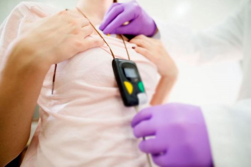
Full dynamic electrocardiogram according to Holter: what is it?
The complete dynamic electrocardiogram according to Holter is a painless and non-invasive test that allows the electrical activity of the heart to be monitored over a 24-hour period, analysing approximately 100,000 beats
The complete dynamic ECG according to Holter allows the electrical activity of the heart to be monitored during the patient’s normal activities
It therefore highlights the correlation between activity and any symptoms and/or changes in the electrocardiogram.
It is a useful test, harmless, repeatable over time, of low cost and with good diagnostic and prognostic power.
Electrocardiogram (ECG): what it is and when it is performed
The electrocardiographic trace, commonly known as ECG, is the easiest and most practical method of observing whether the heart’s electrical activity is normal or whether mechanical or bioelectric pathologies are present.
The ECG electrocardiogram is the graphic recording of the heart’s electrical activity and the changes that occur during cardiac contraction (systole) and relaxation (diastole) of the atria and ventricles during its operation, collected by means of electrodes placed above the surface of the body.
The principle on which it is based is purely physiological: impulses in the myocardium lead to the generation of potential differences, which vary in space and time and are recorded via the electrodes.
It is an absolutely painless test that is the diagnostic method par excellence for all arrhythmias, but also the simplest way of assessing the state of the heart muscle and detecting minor metabolic disorders.
FIRST AID: VISIT THE DMC DINAS MEDICAL CONSULTANTS BOOTH AT EMERGENCY EXPO
Why and when an ECG is used
Thanks to the information provided by the electrocardiogram, it is possible to identify the presence of disturbances in the heart rhythm or in the propagation of the electrical impulse (which causes depolarisation of muscle fibres) but also myocardial alterations resulting from ischaemic suffering (coronary artery disease).
Cardiac problems in which the role of the electrocardiogram is crucial are:
- angina pectoris;
- arrhythmias;
- ischaemic heart disease in its various clinical forms;
- conduction disorders;
- myocardial infarction;
- heart valve diseases;
- heart failure.
The particular morphology of the electrical wave makes it possible to highlight alterations in the diffusion of the stimulus, localised in one of the branches into which the conduction tissue branches at the level of the ventricles.
For example, in myocardial infarction, the ECG is altered both in the acute phase, with the appearance of the characteristic lesion waves, and in the post-acute phase, when the necrosis waves, an expression of the death of a portion of myocardial cells, are evident.
Exercise ECG: when should it be done?
The exercise ECG is a test consisting of continuous recording of the ECG, heart rate and blood pressure during muscular work.
It is generally carried out on a special exercise bike, called a cycle ergometer, or on a treadmill.
These instruments allow gradually increasing efforts to be applied, which can be accurately evaluated in watts.
The exercise electrocardiogram is a very low-risk test that provides extremely important information on cardiac function, especially of a patient following a heart attack.
It makes it possible to determine whether there are still regions of the heart that are poorly perfused, ischaemic and at risk for future events, and thus to draw up a safer prognosis and choose the most suitable treatment.
Electrocardiogram ECG: how to read it?
Reading an Electrocardiogram ECG may at first sight appear to be a difficult task for those who are not medical experts, but in reality, thanks to some simple indications, we can get a general idea and assess the most significant aspects of an ECG by following these guidelines:
P wave: this is the first wave that displays the state of activation/depolarisation of the atria. The size of this wave is usually very small. It measures the time taken by the impulse to propagate to both atria: this can be used to diagnose atrial pathologies such as flutter;
PQ tract: flat and wave-free, it measures the time from the moment the atria begin to activate until the moment the ventricles activate;
QRS complex: this is a set of three waves following one another, which correspond to the depolarisation of the ventricles. These waves give indications of arrhythmias, fibrillations and can also be useful in cases of myocardial infarction;
ST segment: this long ST interval – which follows the S wave and includes the T wave – can detect ischaemic problems, as it represents the period when the ventricles contract and then return to rest;
T wave: represents repolarisation of the ventricles, i.e. the time when the ventricles have finished their activation phase and are ready for a new contraction. It is not always identifiable, as it can also be very small in value. The T wave provides indications of cardiac hypertrophy, myocardial infarction and cardiac ischaemia;
QT interval: this is the representation of the electrical systole, i.e. that period of time in which depolarisation and repolarisation of the ventricles occur. The duration of this interval varies depending on the heart rate.
Why and when to do the dynamic Holter electrocardiogram
In the post-infarction period, the dynamic Holter electrocardiogram provides important indications regarding the occurrence of any dangerous or threatening heart rhythm disturbances, as well as any symptomatic ischaemic episodes, i.e. accompanied by angina pain, silent, not accompanied by pain.
Read Also
Emergency Live Even More…Live: Download The New Free App Of Your Newspaper For IOS And Android
Holter Monitor: How Does It Work And When Is It Needed?
What Is Patient Pressure Management? An Overview
Head Up Tilt Test, How The Test That Investigates The Causes Of Vagal Syncope Works
Cardiac Syncope: What It Is, How It Is Diagnosed And Who It Affects
Holter Blood Pressure: What Is The ABPM (Ambulatory Blood Pressure Monitoring) For?
Sinus Tachycardia: What It Is And How To Treat It

Head Up Tilt Test, How The Test That Investigates The Causes Of Vagal Syncope Works
Aslanger Pattern: Another OMI?
Abdominal Aortic Aneurysm: Epidemiology And Diagnosis
What Is The Difference Between Pacemaker And Subcutaneous Defibrillator?
Heart Disease: What Is Cardiomyopathy?
Inflammations Of The Heart: Myocarditis, Infective Endocarditis And Pericarditis
Heart Murmurs: What It Is And When To Be Concerned
Clinical Review: Acute Respiratory Distress Syndrome
Cardiac Holter, The Characteristics Of The 24-Hour Electrocardiogram
Botallo’s Ductus Arteriosus: Interventional Therapy
Heart Valve Diseases: An Overview
Cardiomyopathies: Types, Diagnosis And Treatment
First Aid And Emergency Interventions: Syncope
Tilt Test: What Does This Test Consist Of?
Cardiac Syncope: What It Is, How It Is Diagnosed And Who It Affects
New Epilepsy Warning Device Could Save Thousands Of Lives
Understanding Seizures And Epilepsy
First Aid And Epilepsy: How To Recognise A Seizure And Help A Patient
Neurology, Difference Between Epilepsy And Syncope
Positive And Negative Lasègue Sign In Semeiotics
Wasserman’s Sign (Inverse Lasègue) Positive In Semeiotics
Positive And Negative Kernig’s Sign: Semeiotics In Meningitis
Lithotomy Position: What It Is, When It Is Used And What Advantages It Brings To Patient Care
Trendelenburg (Anti-Shock) Position: What It Is And When It Is Recommended
Prone, Supine, Lateral Decubitus: Meaning, Position And Injuries
Stretchers In The UK: Which Are The Most Used?
Does The Recovery Position In First Aid Actually Work?
Reverse Trendelenburg Position: What It Is And When It Is Recommended
Drug Therapy For Typical Arrhythmias In Emergency Patients
Canadian Syncope Risk Score – In Case Of Syncope, Patients Are Really In Danger Or Not?
What Is Ischaemic Heart Disease And Possible Treatments
Percutaneous Transluminal Coronary Angioplasty (PTCA): What Is It?
Ischaemic Heart Disease: What Is It?
EMS: Pediatric SVT (Supraventricular Tachycardia) Vs Sinus Tachycardia
Paediatric Toxicological Emergencies: Medical Intervention In Cases Of Paediatric Poisoning
Valvulopathies: Examining Heart Valve Problems