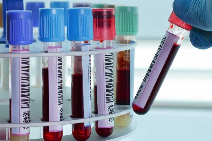
Complete blood count: complete guide to all normal and pathological blood values
The complete blood count is one of the most requested and important blood tests. Blood is made up of a liquid part called plasma and a corpuscular part, made up of cells
The cells divide into red blood cells or erythrocytes, white blood cells or leukocytes, and platelets or thrombocytes.
The blood count, in a single item, contains several measurements
The red blood cells
Red blood cells, or erythrocytes or red blood cells are cells without a nucleus, in the shape of a biconcave disc of 7.3 µ in diameter.
They are produced by cells of the erythroblastic series of the bone marrow.
Red blood cells contain hemoglobin (Hb) which carries oxygen and gives blood its typical red color.
The average values of erythrocytes are 5 million/mm3 in men and 4.5 million/mm3 in women.
The reduction in red blood cells is called anemia.
In clinical practice the hemoglobin value is used; the diagnosis of anemia arises for hemoglobin values lower than 13 g/dL for men and 12 g/dL for women.
Anemia may result from decreased blood cell production in the bone marrow, usually due to deficiency of a key component of erythropoiesis (iron or folic acid or vitamin B12) or from increased destruction of circulating red blood cells (haemolytic anemia) or from loss by hemorrhage.
The symptoms of anemia vary according to the hemoglobin values and the rapidity of onset.
Anemia that sets in rapidly, for example due to hemorrhage or haemolysis, can manifest itself with more serious symptoms even due to modest reductions in hemoglobin, while anemia that sets in over a long period of time can also remain asymptomatic or manifest with mild symptoms even with very low hemoglobin values.
Mild anemia is often asymptomatic. Typical symptoms are tiredness (asthenia), breathlessness (dyspnea) and palpitations, especially during physical activity.
If the anemia is severe, there may also be an increase in heart rate and cardiac output with palpitations (perception of the heartbeat), up to heart failure.
You may have symptoms not directly referable to the cardiovascular system, such as headache (headache), fainting (syncope), ringing in the ears (tinnitus), dizziness, irritability, insomnia and difficulty concentrating.
The increase in red blood cells, even up to 12-15 million per mm3, is called polyglobulia or polycythemia and can be primary (Vaquez’s polycythemia vera) or secondary to environmental stimuli (altitude) or diseases such as cyanogenic congenital heart disease.
Hemoglobin and hematocrit
The hemoglobin value expresses its concentration in whole blood, while the hematocrit is the percentage by volume of whole blood including erythrocytes.
The reduction below the normal value of these two indices indicates the presence of anemia.
The normal range is derived from a Gaussian distribution around the normal mean in a healthy population, with variation based on sex, age, and pregnancy.
Between hemoglobin and hematocrit there is a constant correlation expressed by the formula
Hematocrit = Hemoglobin × 3
Hemoglobin is measured directly while hematocrit is calculated from the number of red blood cells and their mean volume (MCV, see below).
Given this constant correlation, the two values are interchangeable and both can be used for the diagnosis of anemia.
By convention, however, both are reported. The reference values of hemoglobin vary according to the laboratory but in general, values between 14 and 18 g/dL for males and 12 and 16 g/dL for females are considered normal.
A very high hematocrit value in sports could indicate the pharmacological use of erythropoietin, a hormone that physiologically stimulates the bone marrow to produce red blood cells, in order to increase oxygen transport and therefore performance.
In addition to being an incorrect practice, it also exposes the athlete to thrombotic risks due to excessive blood viscosity.
In endurance sports, compared to sedentary sports, the hematocrit can be physiologically normal or slightly decreased precisely to maintain the fluidity of the blood necessary to promote the capillary diffusion of oxygen in the peripheral tissues.
Complete blood count: erythrocyte indices (MCV, MCH, MCHC)
In addition to the red blood cell count, certain parameters, called erythrocyte indices, are evaluated in the blood count, which make it possible to clarify the etiology (the cause) of a possible anemia.
MCV
The MCV or mean corpuscular volume, represents the measure of the mean volume of red blood cells and allows to distinguish between normocytic, microcytic and macrocytic anemia, respectively when the blood cell volume is normal (80-96 fL), decreased (<80 fL) or increased (>96 fL).
The MCV could be altered even in the absence of anemia, for example in the case of alcoholism or certain medications.
The MCV may tend to be higher in the endurance athlete than in the sedentary.
It must be considered that the MCV expresses the average volume of red blood cells and therefore, if conditions favoring both microcytosis and macrocytosis coexist, it could be normal; in this case a peripheral blood smear, i.e. the direct vision of the patient’s blood, will allow us to distinguish the two different cell populations.
The peripheral blood smear also allows you to directly evaluate the morphology of the blood cells. Normally red blood cells are of constant size (7.3 µ) and round in shape.
MCH extension
MCH or mean corpuscular hemoglobin measures the weight of hemoglobin in average red blood cells and typically rises and falls in parallel with the MCV.
MCHC extension
The MCHC or mean corpuscular hemoglobin concentration measures the amount of hemoglobin present in the average red blood cell in relation to its size.
RWD extension
The RDW or erythrocyte volume of distribution expresses the variability in the size of erythrocytes, called anisocytosis.
An increase in this index could precede the change in the MCV and be used together with the latter in the classification of anemias.
Reticulocyte count
Reticulocytes are “young” red blood cells which, unlike mature cells without a nucleus, still contain nuclear genetic material.
The reticulocyte count, which is reported as a percentage of total red blood cells, expresses the ability of the bone marrow to produce red blood cells.
This allows us to make an initial distinction between anemia of reduced production due to bone marrow failure, and anemia due to other causes. In practice, when anemia occurs, the marrow tries to compensate by producing more red blood cells, and consequently the percentage of circulating reticulocytes increases.
White blood cells or leukocytes
White blood cells or leukocytes are divided into neutrophils, monocytes, lymphocytes, eosinophils and basophils.
The single or combined increase or decrease of each of these cells can respectively cause leukocytosis (> 11,000/mm3) i.e. increase, or leukopenia, i.e. decrease in white blood cells.
In addition to counting the various types of white blood cells, in the blood count we find the so-called leukocyte formula, that is, the percentage of each cell type compared to the total.
Neutrophils
The most common cause of leukocytosis is neutrophilia (increase in neutrophils > 7.5×10⁹ cells/L).
In clinical practice, the most common cause of neutrophilia are infections, in particular those of bacterial origin which can induce an increase in neutrophils generally equal to 10-25×109 cells/L.
Some infections, for example pneumococcal pneumonia induce an even more marked increase, while in about 25% of cases of bacterial infections neutrophilia is not found.
Viral infections can cause neutrophilia but are often associated with normal white blood cell counts.
Neutropenia (decreased neutrophils) is the most frequent cause of leukopenia.
The most frequent causes of neutropenia are viral infections and the intake of certain drugs (for example some antibiotics).
Lymphocytes
Lymphocytosis (increase in lymphocytes) can be absolute (normal condition in the first 4 or 5 years of life) or relative, with an increase only of the percentage value in the leukocyte formula.
The most common causes of marked lymphocytosis are viral infections, particularly infectious mononucleosis, acute infectious lymphocytosis, and pertussis among bacterial infections.
Lymphocytosis of various degrees is also observed in leukemias.
Lymphopenia can be found in some lymphomas, and is responsible for the immunosuppression typical of these diseases.
Monocytes
Monocytosis (increased monocytes) is found in hematological disorders (leukemia, lymphoma, multiple myeloma) and infections (tuberculosis, endocarditis, mononucleosis).
Eosinophils
Eosinophilia (increased eosinophils) is typically found in allergies and parasitosis, Hodgkin’s lymphoma, and Loeffler fugax infiltrate.
It can be induced by some drugs.
Eosinopenia (decrease in eosinophils) can be seen in ileotyphoid, myocardial infarction, and some adrenocortical diseases.
Basophils
Basophilia (increased basophils) can be neoplastic, usually very marked, and reactive, of a minor entity resulting from allergic reactions, endocrine disorders, some infections.
The platelets
Platelets play an important role in the processes of blood coagulation and hemostasis.
Normal values are 200,000-300,000 × mm3.
Thrombocytopenia or thrombocytopenia (reduction of platelets), can be manifested, depending on the extent, with bleeding from the mucous membranes, petechiae or ecchymoses.
Thrombocytopenia is distinguished from reduced marrow production or from increased immune and non-immune destruction.
The main causes of thrombocytopenia are autoimmune thrombocytopenic purpura, pregnancy (5% of cases), connective tissue diseases (systemic lupus erythematosus), viral infections (infectious mononucleosis, HIV and cytomegalovirus), radiation therapy, alcohol and some drugs (heparin).
Platelet infections or thrombocytosis (increased platelets) are classified into physiological, caused by physical exercise or stress, reactive, resulting from bleeding, haemolytic anemia, infections or tumors and clonal, in the course of lympho-proliferative diseases.
We speak of thrombocytosis for platelet values higher than 350,000-450,000 / µL.
The most common causes of reactive thrombocytosis are infections, especially bacterial ones, inflammatory diseases (rheumatoid arthritis, polymyalgia rheumatica), liver cirrhosis, iron deficiency anemia, some malignancies.
Read Also
Emergency Live Even More…Live: Download The New Free App Of Your Newspaper For IOS And Android
Iron Deficiency Anaemia: What Foods Are Recommended
What Is The Complete Blood Count (CBC Test)?
Iron, Ferritin And Transferrin: Normal Values
Increased ESR: What Does An Increase In The Patient’s Erythrocyte Sedimentation Rate Tell Us?
Anaemia, Vitamin Deficiency Among Causes
Mediterranean Anaemia: Diagnosis With A Blood Test
Colour Changes In The Urine: When To Consult A Doctor
Why Are There Leukocytes In My Urine?
How Iron Deficiency Anemia (IDA) Is Treated
Mediterranean Anaemia: Diagnosis With A Blood Test
Iron Deficiency Anaemia: What Foods Are Recommended
What Is Albumin And Why Is The Test Performed To Quantify Blood Albumin Values?
What Is Cholesterol And Why Is It Tested To Quantify The Level Of (Total) Cholesterol In The Blood?
Gestational Diabetes, What It Is And How To Deal With It
High Ferritin, Low Ferritin, Normal Values, Significance, Treatment: An Overview
Emergency Medicine: Objectives, Exams, Techniques, Important Concepts


