
Mammography with contrast medium (Contrast-enhanced mammography): when and why to perform CESM
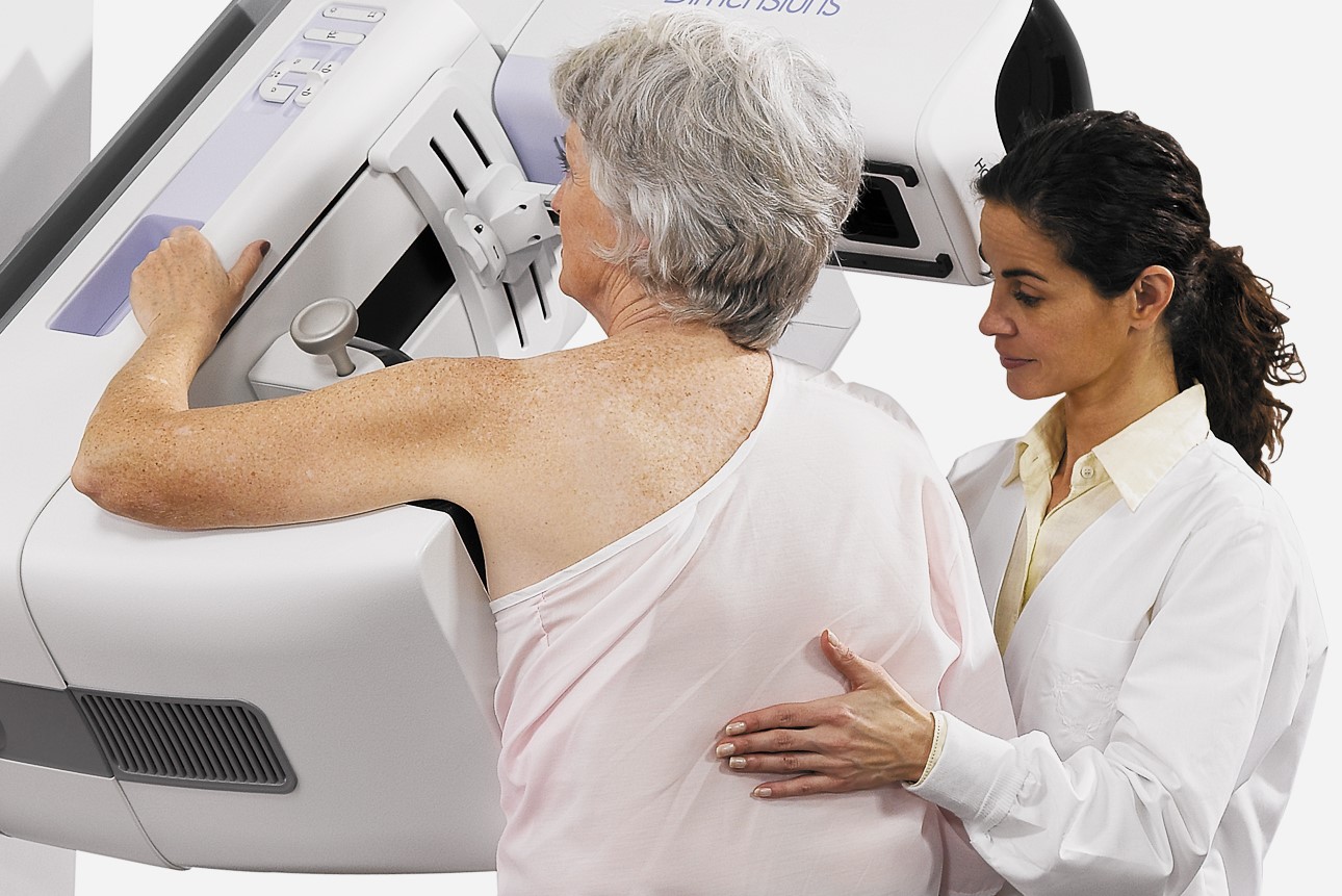
Let’s talk about mammography with contrast medium: the method known as CESM (Contrast Enhanced Spectral Mammography) allows contrastographic assessment of the breast, effectively detecting the presence of neoplasms
Breast cancer is the most frequent neoplasm in the female sex, accounting for about 29% of tumour diagnoses.
Analogue mammography is the reference test for breast screening and has allowed a considerable reduction in mortality (estimated at between 20% and 40%), but it does have some limitations: in particular, its sensitivity is greatly reduced in the presence of dense breasts, as the so-called ‘anatomical noise’ can mask the presence of any lesions.
For this reason, analogue mammography can have a fair number of false negatives, failing to diagnose about 15% of breast tumours, some of them even palpable (hence the need to supplement classical mammography with breast ultrasound and clinical evaluation).
Again, this technique may be less effective at preoperative staging, sometimes failing to diagnose additional multifocal and multicentric foci of disease and thus leading to inadequate treatment.
From analogue to contrast-enhanced mammography
To overcome this problem, analogue mammography has been replaced in recent years by digital mammography which, with its high quality images and better contrast resolution, has made it possible to improve diagnostic accuracy at the screening stage and to develop applications such as tomosynthesis and CESM (Contrast Enhanced Spectral Mammography).
Focusing on the latter method, it should be noted that CESM is the most recent of the diagnostic techniques introduced in the breast field and that the studies published in the international literature demonstrate its high diagnostic performance, thanks to sensitivity, specificity and accuracy values similar to those of contrast-enhanced MRI, which is, however, less specific in investigations in this field.
In addition, CESM is also less expensive and better tolerated by patients
In detail, CESM combines the principles of mammography with the administration of intravenous contrast medium (mdc), which allows, as in MRI, contrastographic assessment of the breast and better localisation of lesions.
The indications for using the CESM test are: presence of doubtful or suspicious lesions, preoperative local staging for breast cancer, differential diagnosis between scar and recurrence, Cup Syndrome (metastatic axillary lymphadenopathy in the absence of a primary breast tumour), patients with absolute or relative contraindications to MRI.
In order to perform the test, the patient must be fasting, have had a blood test confirming that the creatinine value is within the normal range, and have had a pharmacological preparation in case of allergic diathesis, after consultation with our Anaesthesia and Resuscitation specialist.
The good results obtained with CESM encourage further clinical studies to define the role of this method in the breast diagnostic process
Looking at the execution of the test, first of all iodinated mdc is administered, with a dose of about 1.5ml/kg with an automatic injector to ensure a constant flow of 2.5-3 ml/sec.
Two minutes after the administration of mdc, a series of low and high energy images are acquired in rapid succession while the breast remains compressed for the few seconds required to perform the test.
In total, the test lasts less than 10 minutes.
Ultimately, the results of the studies conducted to date have shown that CESM guarantees a high degree of accuracy in the diagnosis of breast cancer and in the evaluation of suspicious lesions, not least because correct size assessment is of fundamental importance in pre-surgical staging for proper preoperative planning and in order to obtain disease-free resection margins, so as to avoid traumatic changes in the body of the woman with carcinoma as much as possible.
Read Also
Emergency Live Even More…Live: Download The New Free App Of Your Newspaper For IOS And Android
Mammography With Tomosynthesis: What It Is And What Advantages It Offers
Pap Test, Or Pap Smear: What It Is And When To Do It
Mammography: A “Life-Saving” Examination: What Is It?
Breast Cancer: Oncoplasty And New Surgical Techniques
Gynaecological Cancers: What To Know To Prevent Them
Ovarian Cancer: Symptoms, Causes And Treatment
What Is Digital Mammography And What Advantages It Has

What Are The Risk Factors For Breast Cancer?
Breast Cancer Women ‘Not Offered Fertility Advice’
Ethiopia, The Minister Of Health Lia Taddesse: Six Centers Against Breast Cancer
Breast Self-Exam: How, When And Why
MRI, Magnetic Resonance Imaging Of The Heart: What Is It And Why Is It Important?
Mammary MRI: What It Is And When It Is Done
Breast Cancer Diagnosis: The Importance Of Periodic Mammography
Mammography: How To Do It And When To Do It
Pap Test: What Is It And When To Do It?
Breast Cancer: For Every Woman And Every Age, The Right Prevention
Transvaginal Ultrasound: How It Works And Why It Is Important
What Is Tomosynthesis (3D Mammography)?
Fusion Prostate Biopsy: How The Examination Is Performed
CT (Computed Axial Tomography): What It Is Used For
What Is An ECG And When To Do An Electrocardiogram
MRI, Magnetic Resonance Imaging Of The Heart: What Is It And Why Is It Important?
Mammary MRI: What It Is And When It Is Done
What Is Needle Aspiration (Or Needle Biopsy Or Biopsy)?
Positron Emission Tomography (PET): What It Is, How It Works And What It Is Used For
CT, MRI And PET Scans: What Are They For?
MRI, Magnetic Resonance Imaging Of The Heart: What Is It And Why Is It Important?
Urethrocistoscopy: What It Is And How Transurethral Cystoscopy Is Performed
What Is Echocolordoppler Of The Supra-Aortic Trunks (Carotids)?