
Mammography: how to do it and when to do it
Mammography is an X-ray test that allows us to examine the breast tissue inside. Like all X-rays, it involves exposing the part under investigation to small doses of ionising radiation, X-rays, capable of detecting pathological changes
We speak of:
- clinical or diagnostic mammography when it is used to diagnose the benign or malignant nature of a lump or any other abnormality of the breast or nipple detected by the doctor or the woman herself.
- screening mammography when the test is carried out on a large scale on a healthy population in order to detect neoplasms in an as-yet unexposed stage.
How to prepare for mammography
Mammography is performed on an outpatient basis and does not require any preparation.
It is advisable not to apply deodorants, lotions and, above all, talcum powder on the day of the test – under the arms or on the breasts – as it can show up on the X-ray image as white dots simulating pathological micro-calcifications.
The test should preferably be performed in the first part of the menstrual cycle when there is less thickening and tension of the mammary gland.
Moreover, at this stage, there is the certainty that the woman is not pregnant.
In fact, although the radiation emitted by the latest generation of mammography machines is very low-dose, a teratogenic risk to the foetus cannot be ruled out, especially in the very first months of pregnancy.
Where and how mammography is performed
Mammography can be performed in hospitals that have appropriate equipment or in health centres that are equipped and in compliance with the regulations on radiology cabinets.
In order to encourage the use of mammography as a screening tool, during breast cancer prevention campaigns, many hospitals, cancer centres and groups of health workers, supported by voluntary associations, perform the test on suitably equipped mobile centres.
The mammograph, also known as a mammography unit, consists of:
- a column with a high-frequency power supply that supports an X-ray tube, where X-rays are produced
- a support base
- a device consisting of a transparent plastic paddle that clamps, compresses and positions the breast in order to obtain images at different angles
- a detection system where the images are imprinted.
Mammography unit
The unit is used exclusively for radiographic tests of the breast, with special accessories that allow only the target part to be exposed to X-rays.
The breast is an organ consisting of fibro- glandular tissue immersed in adipose tissue.
This conformation makes it particularly suitable for radiographic investigation and identification of even millimetric changes in its tissue.
In fact, unlike bones, which absorb most of the radiation and therefore appear white on the X-ray, soft tissues (muscles, fat, organs), which are easily penetrated by X-rays, appear in various shades of grey depending on their consistency: this allows the detection of pathological formations that have a different structure to the surrounding tissue.
With modern digital mammography, the so-called plates – where images used to be imprinted and then printed – are replaced by electronic components that convert X-rays into mammographic images transmitted directly to a computer for reading by the radiologist and for long-term storage.
This system, very similar to digital cameras, allows better quality images to be obtained with a lower dose of radiation.
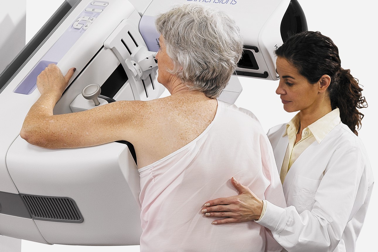
Mammography: how it is performed and how long it lasts
A radiologist places the breast on the special platform of the mammography unit and gradually compresses it with a transparent plastic paddle.
This procedure can cause pain, which, however, only lasts as long as it takes to perform the test.
Compression of the breast is necessary because it smoothes out the thickness of the organ so that it can be viewed in its entirety and results in a lower X-ray dose and higher image quality because a thinner layer of tissue is examined.
In screening mammography, two frames are acquired: one in the head-foot direction and one in the latero-lateral direction.
The screening test lasts a total of 5 to 10 minutes and is conducted by the radiology technician.
Clinical mammography, on the other hand, may require a larger number of projections, including enlarged projections for the study and investigation of details.
It requires the presence of a doctor and may be combined with a breast examination and ultrasound test.
When to perform mammography
Mammography has no particular contraindications.
In women under 40-45 years of age, it may be less readable than ultrasound scans due to the density of the mammary gland.
Clinical mammography is performed whenever, regardless of age, abnormalities of the breast, detected by the doctor or the patient herself, are present in order to assess their nature.
Screening mammography, which is fundamental for early diagnosis because it can reveal breast tissue abnormalities several years before the clinical evaluation, is performed every two years in women aged between 50 and 69, according to the indications of the international guidelines for the prevention of breast cancer, taken up and adopted by the Italian Ministry of Health.
Participation in screening at this frequency and modality reduces mortality by 30%.
Several institutions, especially in the United States, recommend starting screening from the age of 40 onwards on an annual or biannual basis, but there is no agreement on the usefulness of this procedure.
In fact, under the age of 50, the structure of the breast is still very dense, and therefore not easily penetrated by X-rays, resulting in a significant reduction in the diagnostic capacity of the mammography test.
Most studies have not recorded any decrease in mortality in women screened between the ages of 40 and 50.
Longer life spans and continued good health, together with diagnostic efficacy, have led researchers to consider the extension of the screening age up to 74 years to be beneficial.
However, even on the benefits of this strategy there is no convincing data.
Is mammography radiation dangerous?
Exposure to ionising mammographic radiation is not to be considered dangerous.
In particular, digital mammography, compared to traditional analogue mammography, further reduces the amount of X-rays released on the breast.
Scientific studies deny that the radiation doses used in mammography can increase the risk of tumours in the breast and in other districts, even if the test is performed several times in a lifetime.
The limitations of mammography
Screening for breast cancer is now an established practice that has proven effective in reducing mortality from this cancer.
However, the procedure has limitations both by default (false negatives) and by excess (false positives and overdiagnosis).
False negatives
It is estimated that in 20-30% of cases (i.e., 5 out of every 1000 women who have had a mammography performed biannually over a 10-year period) the neoplasm is not detected by the X-ray test.
The causes of the missed diagnosis are to be found in the size and characteristics of the tumour (either too small or poorly defined due to poor contrast), in the structure of the mammary gland that is too dense, in errors of interpretation or in failure of the radiologist to detect the lesion.
Breast implants are an obstacle to accurate mammography readings because, whether silicone or saline implants, they are not transparent to X-rays and can obstruct a clear view of the surrounding tissue, especially if the implant has been placed in front of rather than under the chest muscles.
Overdiagnosis

Mammography may show a malignant lesion but not one that is likely to progress: thus, irrelevant to a woman’s quality of life and life expectancy.
Unfortunately, however, as yet we have no tests that can determine whether the detected tumour will progress or remain indolent, so that there is a possibility, albeit unquantifiable, of diagnosing a lesion – with the consequent in-depth diagnosis and subsequent treatment – that will not develop into a potentially fatal invasive cancer.
Up-to-date research suggests that this risk is lower than the benefits of screening according to the protocol scheme.
False positives
Mammography may show lesions suspicious for cancer that are not confirmed by subsequent tests.
It is estimated that 24% of women (i.e., almost 1 in 4 who have a mammogram every 10 years) receive a diagnosis at least once in a 10-year period that later tests prove to be a false alarm.
Such tests can be invasive, such as a biopsy, and obviously cause concern, but the benefit of ruling out or confirming the presence of a tumour far outweighs any discomfort.
What is the difference between mammography and breast ultrasound?
The difference between ultrasound and mammography lies in the peculiarities of the equipment that lead to different investigation patterns.
The ultrasound scanner uses a probe that emits ultrasound. The sound waves emitted are reflected in different ways on tissues or internal organs and produce different types of echoes, which are then transformed into images on a computer screen.
Mammography uses X-rays that are absorbed by the various tissues according to their consistency
The two systems are therefore complementary.
In younger women, in whom the glandular tissue is denser, ultrasound results offer more information than mammography.
It makes it possible to detect any neoformations within the breast, to distinguish between neoformations with liquid and solid content, and to identify fibroadenomas and areas of fibrocystic mastopathy that are impermeable to mammography testing.
In women over the age of 50, whose breasts have a higher percentage of adipose tissue, mammography is the main indication for the detection of even millimetric tumour lesions.
Read Also
Emergency Live Even More…Live: Download The New Free App Of Your Newspaper For IOS And Android
Breast Cancer: For Every Woman And Every Age, The Right Prevention
Transvaginal Ultrasound: How It Works And Why It Is Important
Pap Test, Or Pap Smear: What It Is And When To Do It
Mammography: A “Life-Saving” Examination: What Is It?
Breast Cancer: Oncoplasty And New Surgical Techniques
Gynaecological Cancers: What To Know To Prevent Them
Ovarian Cancer: Symptoms, Causes And Treatment
What Are The Risk Factors For Breast Cancer?
Breast Cancer Women ‘Not Offered Fertility Advice’
Ethiopia, The Minister Of Health Lia Taddesse: Six Centers Against Breast Cancer
Breast Self-Exam: How, When And Why
Fusion Prostate Biopsy: How The Examination Is Performed
CT (Computed Axial Tomography): What It Is Used For
What Is An ECG And When To Do An Electrocardiogram
MRI, Magnetic Resonance Imaging Of The Heart: What Is It And Why Is It Important?
Mammary MRI: What It Is And When It Is Done
What Is Needle Aspiration (Or Needle Biopsy Or Biopsy)?
Positron Emission Tomography (PET): What It Is, How It Works And What It Is Used For
CT, MRI And PET Scans: What Are They For?
MRI, Magnetic Resonance Imaging Of The Heart: What Is It And Why Is It Important?
Urethrocistoscopy: What It Is And How Transurethral Cystoscopy Is Performed
What Is Echocolordoppler Of The Supra-Aortic Trunks (Carotids)?
Surgery: Neuronavigation And Monitoring Of Brain Function
Robotic Surgery: Benefits And Risks
Refractive Surgery: What Is It For, How Is It Performed And What To Do?
Single Photon Emission Computed Tomography (SPECT): What It Is And When To Perform It
What Is An ECG And When To Do An Electrocardiogram
MRI, Magnetic Resonance Imaging Of The Heart: What Is It And Why Is It Important?
Mammary MRI: What It Is And When It Is Done