
Breast cancer diagnosis: the importance of periodic mammography
Screening mammography is a specific type of breast imaging that uses low-dose x-rays to detect cancer early – before women experience symptoms – when it is most treatable
Tell your doctor about any breast symptoms or problems, prior surgeries, hormone use, whether you have a family or personal history of breast cancer, and if there’s a possibility you are pregnant.
If possible, obtain copies of your prior mammograms and make them available to your radiologist on the day of your exam.
Leave jewelry at home and wear loose, comfortable clothing.
You may be asked to wear a gown.
Don’t wear deodorant, talcum powder or lotion under your arms or on your breasts as these may appear on the mammogram and interfere with correct diagnosis.
What is Mammography?
Mammography is specialized medical imaging that uses a low-dose x-ray system to see inside the breasts.
A mammography exam, called a mammogram, aids in the early detection and diagnosis of breast diseases in women.
An x-ray exam helps doctors diagnose and treat medical conditions.
It exposes you to a small dose of ionizing radiation to produce pictures of the inside of the body.
X-rays are the oldest and most often used form of medical imaging.
Three recent advances in mammography include digital mammography, computer-aided detection and breast tomosynthesis.
Digital mammography, also called full-field digital mammography (FFDM), is a mammography system in which the x-ray film is replaced by electronics that convert x-rays into mammographic pictures of the breast.
These systems are similar to those found in digital cameras and their efficiency enables better pictures with a lower radiation dose.
These images of the breast are transferred to a computer for review by the radiologist and for long term storage.
The patient’s experience during a digital mammogram is similar to having a conventional film mammogram.
Computer-aided detection (CAD) systems search digitized mammographic images for abnormal areas of density, mass, or calcification that may indicate the presence of cancer.
The CAD system highlights these areas on the images, alerting the radiologist to carefully assess this area.
Breast tomosynthesis, also called three-dimensional (3-D) mammography and digital breast tomosynthesis (DBT), is an advanced form of breast imaging where multiple images of the breast from different angles are captured and reconstructed (“synthesized”) into a three-dimensional image set.
In this way, 3-D breast imaging is similar to computed tomography (CT) imaging in which a series of thin “slices” are assembled together to create a 3-D reconstruction of the body.
Although the radiation dose for some breast tomosynthesis systems is slightly higher than the dosage used in standard mammography, it remains within the FDA-approved safe levels for radiation from mammograms.
Some systems have doses very similar to conventional mammography
Large population studies have shown that screening with breast tomosynthesis results in improved breast cancer detection rates and fewer “call-backs,” instances where women are called back from screening for additional testing because of a potentially abnormal finding.
Breast tomosynthesis may also result in:
- earlier detection of small breast cancers that may be hidden on a conventional mammogram
- fewer unnecessary biopsies or additional tests
- greater likelihood of detecting multiple breast tumors
- clearer images of abnormalities within dense breast tissue
- greater accuracy in pinpointing the size, shape and location of breast abnormalities
What are some common uses of the procedure?
Mammograms are used as a screening tool to detect early breast cancer in women experiencing no symptoms.
They can also be used to detect and diagnose breast disease in women experiencing symptoms such as a lump, pain, skin dimpling or nipple discharge.
Screening Mammography
Mammography plays a central part in early detection of breast cancers because it can show changes in the breast years before a patient or physician can feel them.
Current guidelines from the American College of Radiology (ACR) and the National Comprehensive Cancer Network (NCCN) recommend screening mammography every year for women, beginning at age 40.
Research has shown that annual mammograms lead to early detection of breast cancers, when they are most curable and breast-conservation therapies are available.
The ACR and the National Cancer Institute (NCI) also suggest that women who have had breast cancer, and those who are at increased risk due to a family history of breast or ovarian cancer, should seek expert medical advice about whether they should begin screening before age 40 and the need for other types of screening.
If you are at high risk for breast cancer, you may need to obtain a breast MRI in addition to your annual mammogram.
How should I prepare for the mammogram?
Before scheduling a mammogram, the American Cancer Society (ACS) and other specialty organizations recommend that you discuss any new findings or problems in your breasts with your doctor.
In addition, inform your doctor of any prior surgeries, hormone use, and family or personal history of breast cancer.
Do not schedule your mammogram for the week before your menstrual period if your breasts are usually tender during this time.
The best time for a mammogram is one week following your period. Always inform your doctor or x-ray technologist if there is any possibility that you are pregnant.
The ACS also recommends you:
- Do not wear deodorant, talcum powder or lotion under your arms or on your breasts on the day of the exam. These can appear on the mammogram as calcium spots.
- Describe any breast symptoms or problems to the technologist performing the exam.
- Obtain your prior mammograms and make them available to the radiologist if they were done at a different location. This is needed for comparison with your current exam and can often be obtained on a CD.
- Ask when your results will be available; do not assume the results are normal if you do not hear from your doctor or the mammography facility.
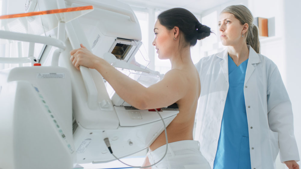
What does the mammography equipment look like?
A mammography unit is a box with a tube that produces x-rays.
The unit is used exclusively for breast x-ray exams and features special accessories to limit x-ray exposure to only the breast.
The unit features a device to hold and compress the breast and position it so the technologist can capture images at different angles.
Breast tomosynthesis is performed using digital mammography units, but not all digital mammography machines are equipped to perform tomosynthesis imaging.
How does the procedure work?
X-rays are a form of radiation like light or radio waves. X-rays pass through most objects, including the body.
The technologist carefully aims the x-ray beam at the area of interest.
The machine produces a small burst of radiation that passes through your body.
The radiation records an image on photographic film or a special detector.
Different parts of the body absorb the x-rays in varying degrees.
Dense bone absorbs much of the radiation while soft tissue (muscle, fat, and organs) allow more of the x-rays to pass through them.
As a result, bones appear white on the x-ray, soft tissue shows up in shades of gray, and air appears black.
Most x-ray images are electronically stored digital files.
Your doctor can easily access these stored images to diagnose and manage your condition.
In conventional film and digital mammography, a stationery x-ray tube captures an image from the side and an image from above the compressed breast.

In breast tomosynthesis, the x-ray tube moves in an arc over the breast, capturing multiple images from different angles.
Mammography is performed on an outpatient basis
During mammography, a specially qualified radiologic technologist will position your breast in the mammography unit.
Your breast will be placed on a special platform and compressed with a clear plastic paddle.
The technologist will gradually compress your breast.
Breast compression is necessary in order to:
- Even out the breast thickness so that all of the tissue can be visualized.
- Spread out the tissue so that small abnormalities are less likely to be hidden by overlying breast tissue.
- Allow the use of a lower x-ray dose since a thinner amount of breast tissue is being imaged.
- Hold the breast still in order to minimize blurring of the image caused by motion.
- Reduce x-ray scatter to increase sharpness of picture.
You will be asked to change positions between images.
The routine views are a top-to-bottom view and an angled side view.
The process will be repeated for the other breast.
Compression is still necessary for tomosynthesis imaging in order to minimize motion, which degrades the images.
During screening breast tomosynthesis, two-dimensional images are also obtained or created from the synthesized 3-D images.
You must hold very still and may need to hold your breath for a few seconds while the technologist takes the x-ray.
This helps reduce the possibility of a blurred image.
The technologist will walk behind a wall or into the next room to activate the x-ray machine.
When the examination is complete, the technologist may ask you to wait until the radiologist confirms they have all the necessary images.
The examination process should take about 30 minutes.
Read Also
Emergency Live Even More…Live: Download The New Free App Of Your Newspaper For IOS And Android
MRI, Magnetic Resonance Imaging Of The Heart: What Is It And Why Is It Important?
Mammary MRI: What It Is And When It Is Done
Mammography: How To Do It And When To Do It
Pap Test: What Is It And When To Do It?
Breast Cancer: For Every Woman And Every Age, The Right Prevention
Transvaginal Ultrasound: How It Works And Why It Is Important
Mammography With Tomosynthesis: What It Is And What Advantages It Offers
Pap Test, Or Pap Smear: What It Is And When To Do It
Mammography: A “Life-Saving” Examination: What Is It?
Breast Cancer: Oncoplasty And New Surgical Techniques
Gynaecological Cancers: What To Know To Prevent Them
Ovarian Cancer: Symptoms, Causes And Treatment
What Is Digital Mammography And What Advantages It Has
What Are The Risk Factors For Breast Cancer?
Breast Cancer Women ‘Not Offered Fertility Advice’
Ethiopia, The Minister Of Health Lia Taddesse: Six Centers Against Breast Cancer
Breast Self-Exam: How, When And Why
Fusion Prostate Biopsy: How The Examination Is Performed
CT (Computed Axial Tomography): What It Is Used For
What Is An ECG And When To Do An Electrocardiogram
MRI, Magnetic Resonance Imaging Of The Heart: What Is It And Why Is It Important?
Mammary MRI: What It Is And When It Is Done
What Is Needle Aspiration (Or Needle Biopsy Or Biopsy)?
Positron Emission Tomography (PET): What It Is, How It Works And What It Is Used For
CT, MRI And PET Scans: What Are They For?
MRI, Magnetic Resonance Imaging Of The Heart: What Is It And Why Is It Important?
Urethrocistoscopy: What It Is And How Transurethral Cystoscopy Is Performed
What Is Echocolordoppler Of The Supra-Aortic Trunks (Carotids)?
Surgery: Neuronavigation And Monitoring Of Brain Function
Robotic Surgery: Benefits And Risks
Refractive Surgery: What Is It For, How Is It Performed And What To Do?
Single Photon Emission Computed Tomography (SPECT): What It Is And When To Perform It
What Is An ECG And When To Do An Electrocardiogram