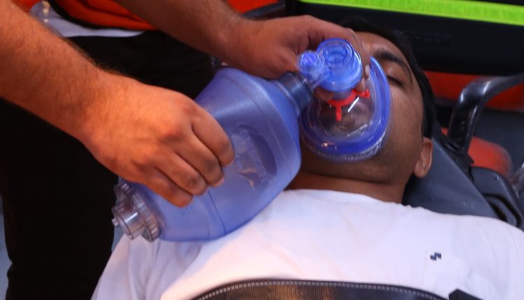
Fires, smoke inhalation and burns: goals of therapy and treatment
Fires are a major cause of injury, death and economic damage. Every year, between 15 and 25 million fires occur in the United States, resulting in approximately 25,000 injuries, 5,000 deaths and $7 to $9 billion in economic damages
The damage induced by smoke inhalation leads to a dramatic worsening of the mortality rate of burn patients: in these cases, smoke inhalation damage is added to burn damage, often with fatal consequences.
This article is devoted to the treatment of burns, with particular reference to pulmonary and systemic damage in burn patients who have inhaled smoke, while dermatological lesions will be discussed in more detail elsewhere.
The objectives of respiratory care in burn patients are to ensure
- airway patency,
- effective ventilation,
- adequate oxygenation,
- the maintenance of acid-base balance,
- the maintenance of cardiovascular stability,
- the prompt treatment of infections.
In some cases, performing an escharotomy is essential to prevent any thoracic scar tissue from impeding chest movement.
The objectives of skin burn treatment consist of
- removal of non-viable skin
- application of medicated bandages with topical antibiotics,
- closure of the wound with temporary skin substitutes and transplantation of skin from healthy areas or cloned specimens to the burned area,
- reducing fluid loss and the risk of infection.
The subject should be given higher than basal caloric quantities to facilitate wound repair and avoid catapolism.
Treatment of burn patients
Burn victims with minor upper airway injuries, or with signs of respiratory obstruction or lung involvement, should be closely monitored.
Oxygen supplementation should be provided, via a nasal cannula, and the patient should be placed in Fowler’s high position to reduce respiratory work.
Bronchospasm should be treated with β-agonists in aerosol (such as orciprenaline or albuterol).
If airway obstruction is anticipated, patency should be ensured with an endotracheal cannula of appropriate calibre.
In general, early tracheostomy is not recommended in burn patients, because this procedure is associated with a higher incidence of infection and increased mortality, although it may be necessary for long-term respiratory care.
It has been observed that early intubation can precipitate transient pulmonary oedema in some patients with inhalation injury.
The application of continuous positive pressure of 5 or 10 cm H2O (CPAP) may help minimise early pulmonary oedema, preserve lung volume, support oedematous airways, optimise the ventilation/perfusion ratio and reduce early mortality.
The administration of systemic corticosteroids for the treatment of oedema is not recommended because of the increased risk of infection.
The treatment of comatose patients should be directed at severe hypoxia and CO poisoning and is based on the administration of oxygen.
The dissociation and elimination of carboxyhaemoglobin are accelerated by the administration of O2 supplements.
Subjects who have inhaled smoke, but only have a slight increase in Hbco (less than 30%) and retain normal cardiopulmonary function, should preferably be treated with 100% O2 delivery, through a tight-fitting, nonrebreathing face mask (which does not allow the freshly exhaled air to be inhaled again), at a flow rate of 15 litres/minute, keeping the reservoir full.
Oxygen therapy should continue until Hbco levels fall below 10%.
A mask CPAP, with 100% O2 administration, may be an appropriate therapy for patients with worsening hypoxaemia and no or only mild thermal injury to the face and upper airway.
Patients with refractory hypoxaemia or inhalation injury associated with coma or cardiopulmonary instability require intubation and respiratory assistance with 100% O2 and should be rapidly referred for hyperbaric oxygen therapy.
The latter treatment rapidly improves oxygen transport and accelerates the process of CO removal from the blood.
Patients who develop early pulmonary oedema, ARDS, or pneumonia often require respiratory assistance with positive end-expiratory pressure (PEEP) in the presence of haemogasanalysis indicative of respiratory failure (PaO2 below 60 mmHg, and/or PaCO2 above 50 mmHg, with pH below 7.25).
PEEP is indicated if PaO2 falls below 60 mmHg and FiO2 demand exceeds 0.60
Ventilatory assistance must often be prolonged, because burn victims generally have an accelerated metabolism, which makes it necessary to increase the respiratory volume per minute to ensure that homeostasis is maintained.
The equipment used must be capable of delivering a high volume/minute (up to 50 litres) while maintaining high peak airway pressures (up to 100 cm H2O) and a stable inspiration/exhalation (I:E) ratio, even when it is necessary to increase pressure values.
Refractory hypoxaemia may respond to pressure-dependent ventilation with an inverted ratio
Adequate pulmonary hygiene is necessary to keep the airways free of sputum.
Passive respiratory physiotherapy helps to mobilise secretions and prevent airway obstruction and atelectasis.
Recent skin grafts do not tolerate percussion and vibration to the chest.
Therapeutic fibrobronchoscopy may be necessary to unblock the airway from thickened secretions.
Careful maintenance of water balance is necessary to minimise the risk of shock, renal failure and pulmonary oedema.
Restoring the patient’s water balance, using Parkland’s formula (4 ml isotonic solution per kg for each percentage point of burnt skin surface area, for 24 hours) and keeping the diuresis between 30 and 50 ml/hour and the central venous pressure between 2 and 6 mmHg, preserves haemodynamic stability.
In patients with inhalation injuries, capillary permeability increases, and monitoring pulmonary arterial pressure is a useful guide to fluid replenishment, in addition to diuresis control.
Fires victims, electrolyte and acid-base balance must be monitored
The hypermetabolic state of the burn patient requires a careful analysis of the nutritional balance, aimed at avoiding catabolism of muscle tissue.
Predictive formulae (such as those of Harris-Benedict and Curreri) have been used to estimate the intensity of metabolism in these patients.
Currently, portable analysers are commercially available that allow for serious indirect calorimetry measurements, which have been shown to provide more accurate estimates of nutritional requirements.
Patients with extensive burns (greater than 50% of the skin surface) are often prescribed diets whose caloric intake is 150% of their resting energy intake to facilitate wound healing and prevent catabolism.
As burns heal, the nutritional intake is progressively reduced to 130% of basal metabolism.
In the case of circumferential burns of the chest, scar tissue may restrict chest wall movement.
The escharotomy (surgical removal of the burnt skin) is performed by making two lateral incisions along the anterior axillary line, starting two centimetres below the clavicle to the ninth to tenth intercostal space, and two other transverse incisions stretched between the ends of the former, so as to delimit a square.
This operation should improve the elasticity of the chest wall and prevent the compressive effect of scar tissue retraction.
Treatment of the burn includes removal of non-viable skin, application of bandages medicated with topical antibiotics, closure of the wound with temporary skin substitutes and transplantation of skin from healthy areas or cloned specimens onto the burned area.
This reduces fluid loss and the risk of infection.
Infections are most often due to coagulase-positive Staphylococcus aureus and gram-negative bacteria such as Klebsiella, Enterobacter, Escherichia coli and Pseudomonas.
Appropriate isolation technique, pressurisation of the environment and air filtration are the cornerstones of defence against infection.
The choice of antibiotic is based on the results of serial cultures performed on material taken from the wound, as well as blood, urine and sputum samples.
Antibiotics should not be administered prophylactically in these patients, due to the ease with which resistant strains can be selected, responsible for infections refractory to therapy.
In individuals who are immobilised for prolonged periods, heparin prophylaxis may help to reduce the risk of pulmonary embolism, and special attention should be paid to preventing the development of pressure sores.
Read Also
Emergency Live Even More…Live: Download The New Free App Of Your Newspaper For IOS And Android
Fires, Smoke Inhalation And Burns: Symptoms, Signs, Rule Of Nine
Calculating The Surface Area Of A Burn: The Rule Of 9 In Infants, Children And Adults
First Aid, Identifyng A Severe Burn
Chemical Burns: First Aid Treatment And Prevention Tips
Electrical Burn: First Aid Treatment And Prevention Tips
6 Facts About Burn Care That Trauma Nurses Should Know
Blast Injuries: How To Intervene On The Patient’s Trauma
What Should Be In A Paediatric First Aid Kit
Compensated, Decompensated And Irreversible Shock: What They Are And What They Determine
Burns, First Aid: How To Intervene, What To Do
First Aid, Treatment For Burns And Scalds
Wound Infections: What Causes Them, What Diseases They Are Associated With
Let’s Talk About Ventilation: What Are The Differences Between NIV, CPAP And BIBAP?
Basic Airway Assessment: An Overview
Respiratory Distress Emergencies: Patient Management And Stabilisation
Respiratory Distress Syndrome (ARDS): Therapy, Mechanical Ventilation, Monitoring
Neonatal Respiratory Distress: Factors To Take Into Account
Signs Of Respiratory Distress In Children: Basics For Parents, Nannies And Teachers
Three Everyday Practices To Keep Your Ventilator Patients Safe
Benefits And Risks Of Prehospital Drug Assisted Airway Management (DAAM)
Clinical Review: Acute Respiratory Distress Syndrome
Stress And Distress During Pregnancy: How To Protect Both Mother And Child
Respiratory Distress: What Are The Signs Of Respiratory Distress In Newborns?
Acute Respiratory Distress Syndrome (ARDS): Guidelines For Patient Management And Treatment
Pathological Anatomy And Pathophysiology: Neurological And Pulmonary Damage From Drowning


