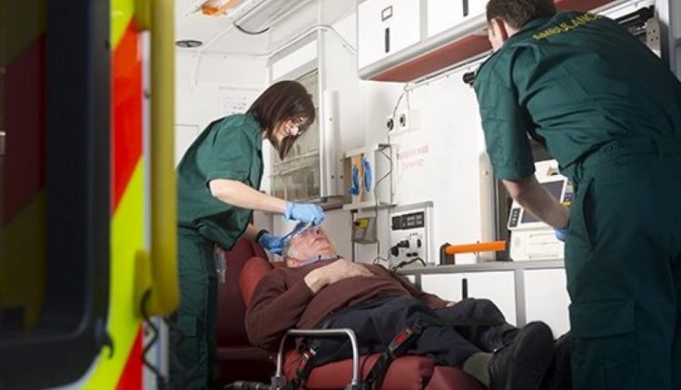
Acute respiratory distress syndrome (ARDS): guidelines for patient management and treatment
The “Acute respiratory distress syndrome” (abbreviated with the acronym ARDS) according to the definition of the WHO (World Health Organization) is a “diffuse damage of the alveolar capillaries causing severe respiratory failure with arterial hypoxemia refractory to the administration of oxygen”
ARDS is therefore a condition, determined by various causes, characterized by the decrease in the concentration of oxygen in the blood, which is refractory to O2 therapy, i.e. this concentration does not rise following the administration of oxygen to the patient.
These pathologies must be treated urgently in intensive care units and, in the most serious cases, can lead to the death of the patient.
ARDS can develop in patients of any age, who already have various types of lung disease, or in subjects with completely normal lung function.
This syndrome is sometimes referred to as adult respiratory distress syndrome, although it can also occur in children.
The less severe form of this syndrome is termed “acute lung injury” (ALI). In the case of a pediatric patient, it is called neonatal respiratory distress syndrome (NRDS).
Conditions and pathologies that predispose to the onset of ARDS are
- drowning;
- suffocation;
- aspiration (inhalation) of food or other foreign material into the lung;
- coronary artery bypass surgery;
- severe burns;
- pulmonary embolism;
- pneumonia;
- pulmonary contusion;
- head trauma;
- traumas of various kinds;
- radiation;
- high altitudes;
- inhalation of toxic gases;
- infections with viruses, bacteria or fungi;
- overdose of drugs or other substances, such as heroin, methadone, propoxyphene, or aspirin;
- sepsis (severe widespread infection);
- shock (prolonged severe arterial hypotension);
- haematological changes;
- obstetric complications (toxemia, amniotic embolism, postpartum endometritis);
- lymphatic obstruction;
- extracorporeal circulation;
- pancreatitis;
- brain stroke;
- seizures;
- transfusions of more than 15 units of blood in a short period of time;
- uremia.
Pathogenesis of ARDS
In ARDS, the small air cavities (alveoli) and pulmonary capillaries are damaged and blood and liquid enter the spaces between the oral cavities and, eventually, inside the cavities themselves.
In ARDS there is an absence or reduction of surfactant (a liquid that coats the inner surface of the alveoli and helps to keep them open), which is responsible for the increased consistency of the lungs typical of ARDS: the surfactant deficiency causes the collapse of many alveoli (atelectasis).
The presence of liquid in the alveoli and their collapse interfere with the transfer of oxygen from the inhaled air to the blood, with a marked reduction in the blood oxygen level.
The transfer of carbon dioxide from blood to exhaled air is less impaired, and blood carbon dioxide levels vary little.
ARDS is characterized by
- acute onset;
- bilateral pulmonary infiltrates suggestive of edema;
- no evidence of left atrial hypertension (PCWP < 18 mmHg);
- PaO2/FiO2 ratio < 200.
- The same criteria, but with a PaO2/FiO2 ratio < 300, define acute lung injury (ALI).
The symptoms of ARDS are
- tachypnea (increased respiratory rate);
- dyspnoea (breathing difficulties with “air hunger”);
- crackles, hissing noises, scattered rales on pulmonary auscultation;
- asthenia (lack of strength);
- general malaise;
- shortness of breath, rapid and shallow;
- respiratory failure;
- cyanosis (appearance of patches or bluish discoloration on the skin);
- possible dysfunction of other organs;
- tachycardia (increased heart rate);
- cardiac arrhythmias;
- mental confusion;
- lethargy;
- hypoxia;
- hypercapnia.
Other symptoms may be present depending on the underlying disease causing ARDS.
ARDS usually develops within 24-48 hours of the trauma or etiological factor, but can occur 4-5 days later.
Diagnosis
Diagnosis and differential diagnosis are based on data collection (medical history), physical examination (especially chest auscultation), and various other laboratory and imaging tests, such as:
- blood count;
- blood gas analysis;
- spirometry;
- lung bronchoscopy with biopsy;
- chest x-ray.
Respiratory insufficiency causes diffuse bilateral accumulations evident on chest x-ray and frequent overlapping infections leading to death in more than 50% of cases.
In the acute phase, the lungs are diffusely enlarged, reddish, congested and heavy, with diffuse alveolar damage (histologically, edema, hyaline membranes, acute inflammation are observed).
The presence of liquid is visible in the spaces that should be filled with air.
In the phase of proliferation and organization, confluent areas of interstitial fibrosis with proliferation of type II pneumocytes appear.
Bacterial superinfections are frequent in fatal cases. Blood gas analysis shows reduced oxygen levels in the blood.
The differential diagnosis includes other respiratory and cardiac disorders and may require other tests, such as an electrocardiogram and cardiac ultrasound.
Neonatal respiratory distress syndrome (NRDS)
NRDS can be observed in 2.5-3% of children admitted to Pediatric Intensive Care Units.
The incidence is inversely proportional to gestational age and birth weight, i.e. the disease is more frequent the more the newborn is premature and underweight.
Neonatal distress is characterized by:
- hypoxia;
- diffuse pulmonary infiltrates on chest X-ray;
- occlusion pressure in pulmonary artery;
- normal heart function;
- cyanosis (bluish color of the skin).
If the respiratory movements are made with the mouth closed, high obstructions must be suspected: the mouth must be opened and the oropharyngeal cavities cleaned of secretions with delicate aspiration.
Most important are the prevention of prematurity (including not performing an unnecessary or untimely caesarean section), the appropriate management of high-risk pregnancy and labour, and the prediction and possible treatment of lung immaturity in utero.
Treatment
Since in 70% of cases the death of the patient occurs NOT for respiratory failure but for other problems related to the underlying cause (mainly multisystem problems that cause renal, hepatic, gastrointestinal or CNS damage or sepsis) the therapy should aim at:
- administer oxygen to counteract hypoxia;
- eliminate the root cause that led to ARDS.
If oxygen given via a facemask or via the nose is not effective in correcting the low blood oxygen levels (which occurs often), or if very large doses of inspired oxygen are required, ventilation should be used. mechanical: a special instrument delivers oxygen-rich air under pressure with a tube which, through the mouth, is introduced into the trachea.
In ARDS patients, the ventilator inputs
- air at increased pressure during inspiration;
- air at lower pressure during exhalation (defined as positive end-expiratory pressure) which helps to keep the alveoli open during the end-expiratory phase.
Treatment takes place in the intensive care unit
The administration of O2 proves to be useful only in the initial stages of the syndrome, however it does not bring benefits on the prognosis.
Endotracheal instillation of multiple doses of exogenous surfactant in low-weight infants requiring 30% oxygen and assisted ventilation: survival is increased, but does not significantly reduce the incidence of chronic lung disease.
Suspicion of ARDS: what to do?
If you suspect ARDS, do not wait any longer and take the person to the Emergency Department, or contact the Single Emergency Number: 112.
Prognosis and mortality
Without effective and timely treatment, ARDS unfortunately causes death in 90% of patients, however, with adequate treatment, approximately 75% of patients survive.
Factors affecting the prognosis are:
- age of the patient;
- general health conditions of the patient;
- comorbidity (presence of other pathologies such as arterial hypertension, obesity, diabetes mellitus, severe lung disease);
- ability to respond to treatment;
- cigarette smoke;
- speed of diagnosis and intervention;
- skill of the healthcare staff.
Patients who respond quickly to treatment are those most likely not only to survive, but also to have little or no long-term lung damage.
Patients who do not respond rapidly to treatment, require long-term ventilator assistance, and are elderly/debilitated are at greatest risk of lung scarring and death.
Scarring can alter lung function, a fact that appears evident with dyspnoea and easy fatigue under effort (in less serious cases) or even at rest (in more serious cases).
Many patients with chronic damage may experience significant weight loss (decrease in body weight) and muscle tone (decrease in % of lean mass) during illness.
Rehabilitation in special specialized rehabilitation centers can be extremely useful for regaining strength and independence during convalescence.
Read Also
Emergency Live Even More…Live: Download The New Free App Of Your Newspaper For IOS And Android
Basic Airway Assessment: An Overview
Respiratory Distress Emergencies: Patient Management And Stabilisation
Respiratory Distress Syndrome (ARDS): Therapy, Mechanical Ventilation, Monitoring
Neonatal Respiratory Distress: Factors To Take Into Account
Signs Of Respiratory Distress In Children: Basics For Parents, Nannies And Teachers
Three Everyday Practices To Keep Your Ventilator Patients Safe
Benefits And Risks Of Prehospital Drug Assisted Airway Management (DAAM)
Clinical Review: Acute Respiratory Distress Syndrome
Stress And Distress During Pregnancy: How To Protect Both Mother And Child
Respiratory Distress: What Are The Signs Of Respiratory Distress In Newborns?
Sepsis: Survey Reveals The Common Killer Most Australians Have Never Heard Of
Sepsis, Why An Infection Is A Danger And A Threat To The Heart
5 Types Of First Aid Shocks (Symptoms And Treatment For Shock)
Obstructive Sleep Apnoea: What It Is And How To Treat It
Obstructive Sleep Apnoea: Symptoms And Treatment For Obstructive Sleep Apnoea
Our respiratory system: a virtual tour inside our body
Tracheostomy during intubation in COVID-19 patients: a survey on current clinical practice
FDA approves Recarbio to treat hospital-acquired and ventilator-associated bacterial pneumonia


