
Spontaneous pneumothorax: discussing the collapse of the lungs
Collapse of the lungs due to air entering the space enclosed by the pleurae. If it is not enough to administer oxygen to relieve laboured breathing and drugs to control pain, it will be necessary to remove the air
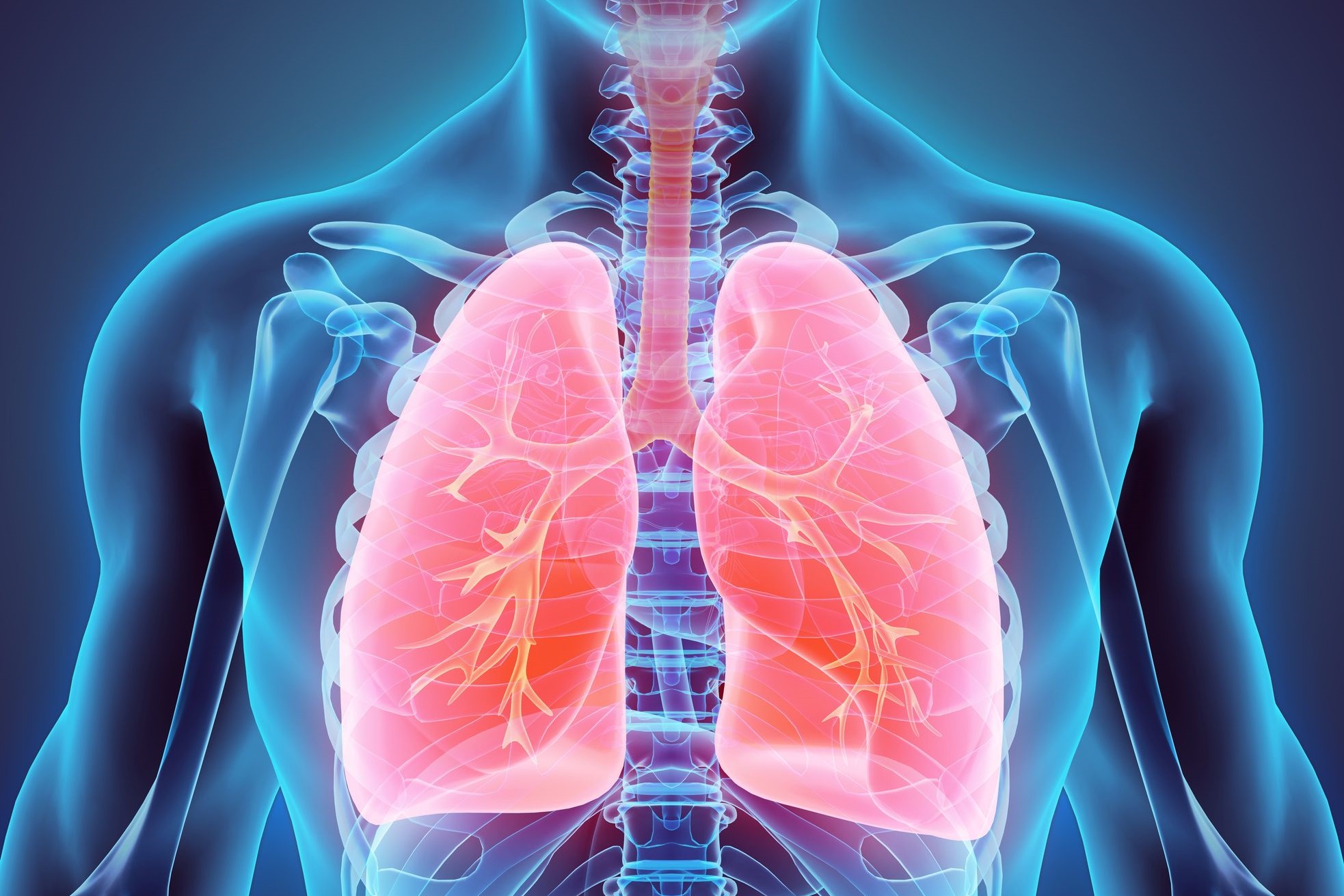
Spontaneous pneumothorax is the collapse of one or both lungs caused by air entering the pleural cavity
The pleura is a thin membrane made up of two ‘sheets’ that line each lung and the inside of the chest.
The pleural cavity is the space between the two leaflets of the pleura and it is here that the abnormal entry of air that causes pneumothorax occurs.
Spontaneous pneumothorax is quite rare in paediatric patients
However, if it occurs once, it is very likely that there will be a relapse.
Spontaneous pneumothorax may have no apparent cause.
Sometimes, it may occur as a result of a child’s lung disease.
The most frequent is an asthmatic condition that is not well controlled.
Very often, pneumothorax is caused by trauma to the chest.
Primary spontaneous pneumothorax is not caused by a lung disease and its causes are essentially unknown
In contrast, in secondary spontaneous pneumothorax, the collapse of the lung is caused by a lung disease.
The most common symptom is a sudden pain in the chest, which becomes more intense during breathing.
Other symptoms are coughing, shoulder pain or a sharp pain between the shoulder blades.
In most cases, these symptoms do not last long and very rarely worsen.
The diagnosis is based on a careful collection of the child’s medical history and an equally careful examination.
Spontaneous pneumothorax is often diagnosed by a chest X-ray in the emergency room.
If the doctor needs more detailed images of the lung, in some cases he or she may also request a computed tomography (CT) scan.
With the CT scan, it will be possible to identify and measure ‘blebs’ (or bullae), which are small air-filled bubbles that can form on the surface of the lung.
Spontaneous pneumothorax can often be caused by the rupture of these bullae, which release air into the pleural cavity.
Treatment is not always necessary immediately after a spontaneous pneumothorax
Young patients are admitted for close observation.
Sometimes it is sufficient to administer oxygen to relieve laboured breathing (dyspnoea) and drugs to control pain.
If, on the other hand, there is a risk that the lung will be excessively compressed by air escaping into the pleural cavity or that spontaneous pneumothorax will impair breathing, it will be necessary to remove the air.
This procedure can be performed by sucking the air out with a needle or by inserting a tube into the chest to let the air escape from the pleural cavity.

When a relapse occurs, it may be necessary to perform an operation to ‘glue’ the pleura and prevent the lung from collapsing again.
Another procedure that can be used is videothoracoscopy, which consists of a relatively non-invasive laparoscopy performed under general anaesthesia.
Videothoracoscopy is performed using a thin, tube-like instrument with a camera at the end.
This tube is inserted into the chest through a small incision and allows air bubbles to be visualised and removed.
Open thoracotomy, i.e. surgical incision of the chest, is less commonly used today for removing bubbles.
The prognosis is usually good and, with the right treatment, the pneumothorax tends to resolve quickly
When the child is discharged, it is recommended that the family take him or her immediately to the emergency room if chest pain or difficulty breathing reappears.
These symptoms may indicate a relapse of the pneumothorax.
Read Also:
Emergency Live Even More…Live: Download The New Free App Of Your Newspaper For IOS And Android
Pulmonary Emphysema: Symptoms, Diagnosis And Treatment
Airway Management After A Road Accident: An Overview
Tracheal Intubation: When, How And Why To Create An Artificial Airway For The Patient
What Is Transient Tachypnoea Of The Newborn, Or Neonatal Wet Lung Syndrome?
Traumatic Pneumothorax: Symptoms, Diagnosis And Treatment
Diagnosis Of Tension Pneumothorax In The Field: Suction Or Blowing?
Pneumothorax And Pneumomediastinum: Rescuing The Patient With Pulmonary Barotrauma
Cervical Collar In Trauma Patients In Emergency Medicine: When To Use It, Why It Is Important
KED Extrication Device For Trauma Extraction: What It Is And How To Use It
ABC, ABCD And ABCDE Rule In Emergency Medicine: What The Rescuer Must Do
Multiple Rib Fracture, Flail Chest (Rib Volet) And Pneumothorax: An Overview
Primary, Secondary And Hypertensive Spontaneous Pneumothorax: Causes, Symptoms, Treatment
Pneumothorax And Haemothorax: Trauma To The Thoracic Cavity And Its Consequences