
Focal shock waves: what they are and when they are used
Focal shock waves, introduced into medicine in the early 1990s to treat kidney stones (urological lithotripsy), have also been used for more than a decade to treat many musculoskeletal disorders (mainly tendons and bone)
A non-invasive method, shock waves are in many cases a valid therapeutic option for treating many diseases, even in the acute phase, thanks to their beneficial anti-inflammatory, pain-relieving and ‘anti-oedema’ (i.e. to combat ‘swelling’) properties, as well as to stimulate tissue repair.
More recently, in fact, they have also proved effective in the field of skin regeneration, accelerating the healing process of sores, ulcers and ‘difficult’ wounds of various origins, including post-traumatic.
What are shock waves?
They are acoustic waves (sound impulses of a mechanical nature), produced by special generators (lithotriptors), which are then able to propagate in the tissues, in a rapid and repeated sequence.
They are characterised by a particular waveform (first phase of positive pressure, followed by an equally rapid phase, less extensive, of negative pressure), which differentiates them from ultrasounds and which, as a whole, is responsible for the positive biological effects applicable in the therapeutic field.
At a microscopic level, stimulation with shock waves is comparable to a sort of deep ‘micro-massage’ of the tissues and cells, capable of inducing the latter to react positively, with the production of substances with anti-inflammatory action and growth factors, which stimulate the regeneration of the tissues themselves, starting from the stem cells.
This type of mechanical stimulation can in many cases also be successfully applied (in association with other codified therapies) to reduce muscular hypertonus in conditions of spasticity of various origins, both in the lower and upper limbs, although the mechanism of action is still partly unknown.
Thanks to these basic biological effects, for more than a decade the use of shock waves has been widely spread from the urological field to the orthopaedic, physiatric and rehabilitation fields, but with substantial differences linked to the fact that they act on living tissues and not on non-viable calcific concretions (such as stones).
Well-tolerated, non-invasive, repeatable and highly effective clinically, focal shock waves, in certain appropriately selected cases, also prove to be an alternative to surgery, or a solution for treating the after-effects of trauma or surgery itself.
Shock waves also:
- can act synergistically (i.e. reinforce) with other therapies, or even enhance and accelerate the expected results of surgery;
- shockwave treatment carried out in the first instance does not preclude the possibility of subsequently intervening with other therapeutic solutions (e.g. surgery).
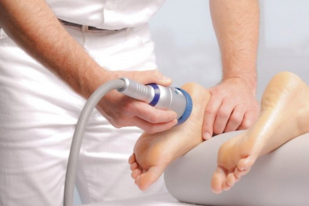
The therapy is carried out in different ways depending on whether the patient is suffering from bone pathologies, tendon and muscle pathologies, spasticity or skin pathologies.
The duration of each session can vary from 10-15 minutes in the case of ‘soft’ tissue applications (tendons, muscles and skin), to longer (up to an hour) for bone treatments.
The patient is generally placed in a supine position on the couch or in a seated position.
Throughout the duration of the therapy the patient is under constant and direct medical supervision, so that the level of energy is also modified according to the patient’s sensitivity.
Shock waves are safe and effective
Treatment with focal shock waves is a non-invasive, outpatient method that is safe and proven to be effective.
The therapy has virtually no clinically relevant side effects and is well tolerated (if correctly performed) and repeatable.
The benefits for the patient are now internationally recognised and have been proven by about 15 years of experience in daily clinical practice.
Follow up with treatment
The treatment may, in some cases, have an immediate pain-relieving effect, but this is not the rule.
Generally, the benefits occur progressively, over weeks.
In order to assess the effectiveness of the treatment correctly, a follow-up period of about 2-3 months is advisable.
During this period, refraining from sporting activity and resting are recommended.
In addition, in the case of treatments carried out for bone consolidation problems (e.g. in pseudarthrosis), since mechanical stability is essential for healing, a brace to immobilize the limb or the use of crutches may be prescribed.
Treatment with focal shock waves is effective in treating many diseases of the bones and ‘soft’ tissues (tendons, ligaments), thanks to its beneficial anti-inflammatory, pain-relieving and ‘anti-oedema’ (i.e. to combat ‘swelling’) properties, as well as stimulating tissue repair:

- shock wave treatment against calcifications
- modulation of shock waves for inflammatory diseases in the acute phase (i.e. more recent onset and already very painful in themselves)
- wave treatment for tissue regeneration (for treating skin ulcers of various origins and related diseases)
- shock wave treatment for treating fractures.
Are shock waves painful or dangerous?
NO, if performed according to codified treatment protocols and with suitable equipment, they are generally well tolerated.
Especially when it comes to treatments for “soft” tissue pathologies (tendons and ligaments).
In some cases, if the patient feels a slight discomfort, the doctor can adjust the intensity of the energy and the number of strokes applied to ensure that the treatment is better tolerated and still effective.
In addition, some treatment protocols provide for a progressive increase in the energy applied, so that the patient can adapt without too much difficulty.
In the case of treatments on the bone, where higher energies are applied for a longer period of time, the pain may be more intense and local anaesthesia may be required.
As this is a non-invasive therapy, it is safe and almost free of major side effects.
In general, they may occur after the application of high energy:
- small haematomas, petechiae and superficial and short-lived bruising;
- temporary awakening of pain symptoms. The flare-up of pain after shock wave treatment should not be interpreted as an adverse or negative event, but as a possible positive response to mechanical stimulation of the tissues.
Focal shock wave treatment is a so-called “manu-medical” therapy, i.e. performed by a physician with specific expertise in the field
Basically, there are no “cons”: like all “biological” treatments, which imply a response from the tissues, the result (especially for bone and, to a lesser extent, skin regeneration) is not immediate, but manifests itself over the months following the end of the treatment.
However, it should be pointed out that shock waves do not replace surgical treatment in all cases.
Do shock waves have contraindications?
The following contraindications, divided into absolute and relative, are currently recognised.
Absolute contraindications are
- the presence of delicate and sensitive structures, such as the encephalon, spinal cord and gonads in the focal field;
- the presence of tumour pathologies and thrombophlebitis where shock waves should be applied;
- pregnancy
- the presence of hollow organs (e.g. lung and intestines) in the focal field (tissue damage may occur when the sound wave passes from solid to gas).
Relative contraindications are considered
- the presence of Pace Makers or electro-stimulators of different origin (in particular for patients with Pace Makers, attention must be paid to the type of generator to be used);
- the proximity of cartilage which is still in the growth phase (in reality, this is now considered more of a precaution than a real contraindication, since numerous experimental studies have demonstrated the absence of harmful effects)
- Diseases or alterations in blood coagulation (coagulopathies with a tendency to bleed): in such cases, the doctor will assess the suitability or otherwise of the treatment for each individual patient, and possibly also the type of instrumentation to be used.
Read Also:
Emergency Live Even More…Live: Download The New Free App Of Your Newspaper For IOS And Android
Pain Management In The Paediatric Patient: How To Approach The Injured Or Aching Children?
Pericarditis In Children: Peculiarities And Differences From That Of Adults
In-Hospital Cardiac Arrest: Mechanical Chest Compression Devices May Improve Patient Outcome
Chest Pain In Children: How To Assess It, What Causes It
Pain Perception In Children: Analgesic Therapy In Paediatrics