
What are myomas? An overview of this benign tumour of the female pelvis
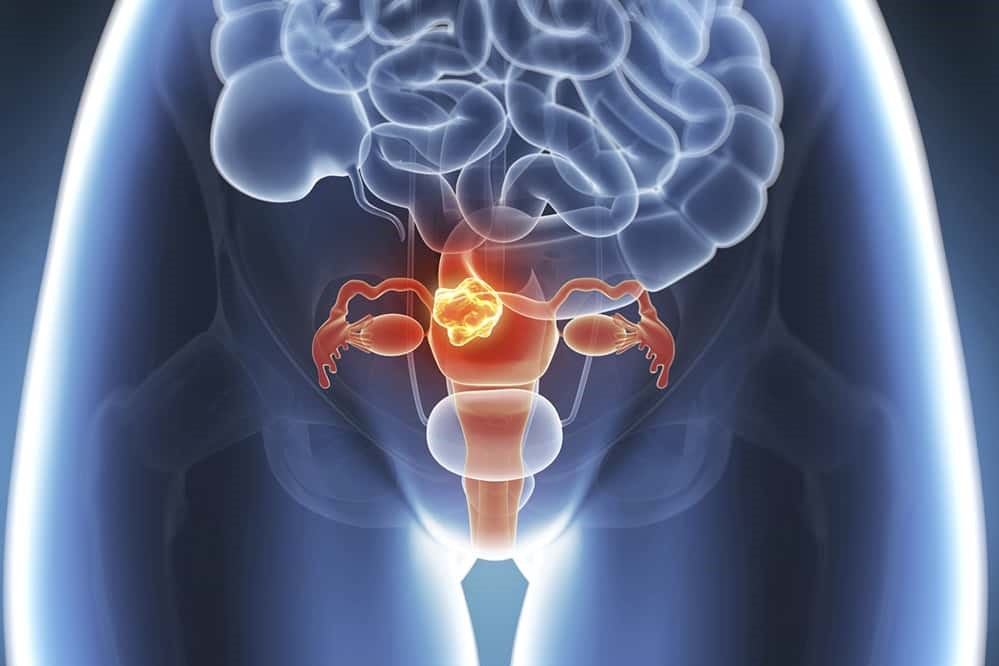
Uterine myomas, or fibroids, are the most common benign tumour of the female pelvis, being found in more than 35% of women over the age of 30
In fact, only 1-4 out of 1000 patients undergoing surgery for myomas have a histological diagnosis of a malignant tumour (leiomyosarcoma).
Myomas, the risk factors
Risk factors for the development of myomas are age, prolonged exposure to endogenous oestrogen (early menarche), family history of fibroids, ethnicity, obesity, nulliparity, and diet.
Myomas are often asymptomatic, but in 40% of cases their presence is the cause of complaints (heavy or short periods, abdominal distension, pelvic pain, repeated miscarriages or infertility) that compromise a woman’s health and quality of life.
Treatment is only necessary when fibroids are symptomatic, and the most frequently used therapies are surgical, consisting of removing the whole uterus (hysterectomy) or individual fibroids (myomectomy).
In recent decades, however, the therapeutic choice for women with symptomatic fibroids has expanded significantly with the advent of an effective non-surgical alternative, uterine artery embolisation.
Myomas and fertility
The presence of submucosal myomas that distort the uterine cavity decreases fertility: a meta-analysis of the literature has shown that this type of myoma reduces the chances of pregnancy by 70%.
Surgical removal of myomas allows normal fertility to be restored.
Submucosal myomas that deform the uterine cavity lead to repeated miscarriages, probably through alterations in the blood vessels that lead to a reduction in the supply of oxygen and nutrients to the endometrium, thus hindering the implantation and development of the embryo.
In addition, they may lead to infertility by other mechanisms such as obstruction of the intrauterine portion of the fallopian tubes or local production of biological factors that interfere with embryo transport at the tubal level.
In contrast, neither intramural nor subserous myomas appear to alter female fertility and their removal does not increase fertility.
Myomas and pregnancy
Pregnancy has variable and unpredictable effects on the growth of myomas.
This variability probably depends on individual differences in genetics, in circulating growth factors, and in the levels of oestrogen and progesterone receptors at the level of the myomas.
An increase in the volume of myomas is present in 30-35% of pregnant women, and this increase occurs predominantly in the first trimester of pregnancy.
In 5-9% of pregnant women with myomas, ultrasound scans demonstrate a process of myoma colliquation.
This phenomenon is a consequence of the rapid growth of the pregnant uterus, which reduces the blood supply to the myomas.
From a clinical point of view, the colliquation of myomas can cause the appearance of abdominal pain requiring hospitalisation and medical therapy (painkillers, antibiotics).
A recent study (Qidwai 2006) compared the outcome of pregnancy in 401 women with ultrasound-tested myomas versus 15104 pregnant women without myomas.

Pregnant women with myomas showed an increase in preterm deliveries (19% vs. 12%), placenta previa (3.5% vs. 1.8%), postpartum haemorrhage (8.3% vs. 2.9%) and the number of caesarean sections (49.1% vs. 21.4%).
Medical treatment
GnRH analogues (which create a state of pharmacological menopause) decrease the volume of uterine, myomas by decreasing oestrogen and progesterone levels.
However, these benefits are temporary and limited to the time of amenorrhoea caused by the analogues.
Upon discontinuation of treatment, the cycle returns after 4-8 weeks, and uterine volume returns to pre-treatment levels in 4-6 months.
Side effects are present in 95% of patients treated with analogues: about 80% of patients have hot flushes, about 30% vaginal dryness, about 55% headache.
The hypoestrogenic state induced by the analogues also leads to a significant loss of bone mass after 6 months of therapy.
RU-486 blocks progesterone receptors and reduces uterine volume, but causes endometrial hyperplasia in 30% of cases.
Read Also:
Emergency Live Even More…Live: Download The New Free App Of Your Newspaper For IOS And Android
Polycystic Ovary Syndrome: Signs, Symptoms And Treatment
Total And Operative Hysterectomy: What They Are, What They Involve
The Symptoms, Diagnosis And Treatment Of Bladder Cancer
Radiotherapy: What It Is Used For And What The Effects Are
Pap Test, Or Pap Smear: What It Is And When To Do It
Drugs Used In Obstetric Emergencies To Modify Uterine Contractions
Polycystic Ovary Syndrome (PCOS): What Are The Symptoms And How To Treat It