
Congenital or acquired malformations: pes cavus
Pes cavus is one of the most common malformations. Those who suffer from it have a more accentuated medial plantar arch, and therefore higher, than it should
The opposite situation is flat foot, a problem characterized by the flattened plantar vault and valgus-pronation of the heel.
Flat feet, common between 10 months and 3-4 years and destined to resolve spontaneously generally within 6-7 years, when it persists can favor the development of problems at the level of the ankles and knees.
Similarly, pes cavus can create a series of postural problems that can cause pain, as the foot does not rest on the ground as it should.
The excessive curvature of the plantar arch, however, is not the only characteristic of this condition: those who suffer from it also have a heel that turns inward and the first metatarsal is lowered.
Congenital or acquired, pes cavus can be classified according to severity and require conservative or surgical intervention.
Flat foot: what it is and how to recognize it
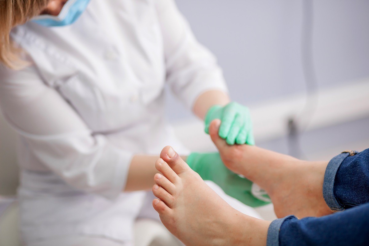
Anatomical deformity, the cavus foot is recognizable on objective investigation.
In fact, those who suffer from it have a higher than normal plantar arch and, when they put their foot on the ground, they are unable to place their toes, heel and part of the sole of the foot at the same time.
The only structures that come into contact with the ground are the heel and toes, while the central part remains “raised”.
In physiological conditions, the foot is simultaneously hollow and flat: it flattens when it is in support (acting as a shock absorber) and the plantar arch raises when pushing.
The hollow foot, therefore, works well in the push phase but does not cushion the step when it rests on the ground
It goes without saying that anyone with this anomaly has a problem in the distribution of body weight.
Instead of weighing on the whole foot, it only weighs on the areas that rest on the ground.
It is a malformation due to two different conditions, which occur simultaneously: the internal structures that form the medial plantar arch rise in an unnatural way, and the anterior area (especially that of the big toe) curves downwards.
Often, tendon and muscle problems also arise.
Pes cavus has several causes, which can be congenital, adaptive, or idiopathic
In the first case the characteristic has been present since birth and genetic factors are involved, in the second case triggering factors are recognizable and in the third it originates for unknown reasons.
Adaptive causes include neurological disease (in 70% of cases), skeletal causes, and trauma.
The neurological diseases that can lead to the origin of the hollow foot are:
- Charcot-Marie-Tooth syndrome, an inherited neuropathy in which the muscles of the lower legs weaken and atrophy;
- Friedreich’s ataxia, hereditary neurodegenerative disease, characterized by progressive ataxia of gait;
- spina bifida, a malformation due to incomplete closure of one or more vertebrae;
- hereditary neuropathy, characterized by sensory loss of small and large nerve fibers;
- the stroke;
- poliomyelitis;
- spinal tumors;
- brain tumors;
- spastic paralysis;
- spinal injuries;
- syringomyelia, a neurological disease characterized by the formation of fluid-filled cysts in the spinal cord;
- muscular dystrophy;
- gout.
Cavus foot can also originate due to some skeletal alterations, which affect the forefoot (forefoot-driven cavus foot) or the hindfoot (hindfoot-driven cavus foot).
In the first case there is a plantar flexion of the first metatarsal, with the foot tending to be hollow and with the hindfoot tending towards supination in response.
In the second case, in addition to the plantar flexion of the first metatarsal, there is an autonomous supination of the hindfoot.
Finally, trauma (to the heel, foot, ankle) or tendon injury (typically, Achilles tendon injury) can cause pes cavus.
The patient generally feels a reduced ability to move the joints, and the disharmonious wear of the footwear shows that there is an asymmetry in the distribution of body weight.
Often, in the case of post-traumatic pes cavus, there is also arthrosis of the ankle (the cartilage tissue is increasingly reduced and, to react to friction, the bone produces osteophytes).
Much more common among women, cavus foot can finally be caused by the prolonged use of heeled shoes, which force the foot into an unnatural position and curvature.
In many cases, the pes cavus is visibly present without symptoms
When symptoms are present, they usually consist of:
- pain in the ankle and foot, especially on the sides and in the metatarsal area (part of the foot skeleton made up of five long, thin bones arranged in parallel);
- unstable ankles, which easily incur sprains;
- difficulty standing upright for long periods, walking or running long distances;
- feeling of stiffness in the feet and ankles;
- claw (or hook) fingers: the phalanges are curved downwards in the central and distal joints, and the fingers bend downwards due to the damage;
- frequent manifestation of calluses on the heels, metatarsus and sides of the foot.
More serious symptoms of pes cavus may consist of peroneal tendonitis (inflammation of the peroneal tendon), Achilles tendon rupture, plantar fasciitis (inflammation and pain in the arched ligament that connects the heel with the base of the toes) and ankle impingement (pain in the front of the ankle caused by impact between two fibrous or skeletal structures).
The diagnosis
If you feel particularly intense pain in your feet and ankles, or a sense of weakness, if you often sprain or have hooked fingers, it is advisable to book a visit to a specialist (orthopaedic or podiatrist).
In most cases, a physical examination and history are sufficient for the diagnosis.
The doctor listens to the patient’s symptoms, inquires about his medical and family history, observes the foot at rest and during walking.
If he deems it appropriate, he can prescribe an X-ray to have a clear idea of the anatomy of the patient’s feet, or an MRI to check the state of the periarticular soft structures.
Sometimes, magnetic resonance imaging of the brain and spinal cord is helpful in investigating whether a neurologic disorder is present.
A final test that can be prescribed is electromyography, to evaluate the conditions of the muscles and the nervous structures that innervate them.
In any case, these are minimally invasive methods and almost totally free of side effects.
The type of treatment depends on what causes pes cavus and how severe the condition is
- It is above all the severity of the symptoms that determines the choice of a conservative or surgical approach.
- In the case of forefoot-driven cavus, in general, a custom-made insole is prescribed to be inserted inside the shoes, useful for improving the impact on the ground and for correcting the distribution of body weight on the foot.
- In the case of hindfoot-driven cavus, on the other hand, the arch support does not give long-term benefits.
Conservative treatment is also indicated for those suffering from neurological hollow foot, due to the ability of the brace to facilitate correct walking.
Other conservative therapies consist of physiotherapy (aimed at improving walking and running and indicated above all for sportsmen), in the prescription of pain-relieving drugs, in the use of shoes designed for those with this genetic anomaly, and in rest from sports activities in the phases in which the pain is sharp.
There are cases, however, where surgical therapy is the only option.
This is necessary to relieve pain, to eliminate or reduce deformity and to avoid otherwise frequent ankle sprains.
For those with forefoot-driven, the most suitable surgical therapy is the osteotomy (and therefore the removal of a bone portion) of the first metatarsal; those with hindfoot-driven, on the other hand, need multiple osteotomies (of the heel, of the first metatarsal).
Other practicable operations are the surgical lengthening of the Achilles tendon, surgical distension of the plantar fascia, tendon transfers and arthrodesis (surgical operations that transform a joint from mobile to static).
Read Also
Emergency Live Even More…Live: Download The New Free App Of Your Newspaper For IOS And Android
Foot Deformities: Metatarsus Adductus Or Metatarsus Varus
Foot Arthrosis: Symptoms, Causes And Treatment
Pain In The Sole Of The Foot: It Could Be Metatarsalgia
Orthopaedics: What Is Hammer Toe?
Hollow Foot: What It Is And How To Recognise It
Occupational (And Non-Occupational) Diseases: Shock Waves For The Treatment Of Plantar Fasciitis
Flat Feet In Children: How To Recognise Them And What To Do About It
Swollen Feet, A Trivial Symptom? No, And Here’s What Serious Diseases They May Be Associated With
Diabetic Foot: Symptoms, Treatment And Prevention
Congenital Malformations: The Discoid Meniscus In Children And Adolescents
Foot Sprain: What It Is, How To Intervene