
How to recognise hip dysplasia?
Congenital hip dysplasia is one of the most common orthopaedic malformations, if not the most common, found in paediatric children
In fact, it is estimated to affect 1-2 infants (mostly girls) per 1000 and, if not properly diagnosed, can also lead to serious consequences such as dislocation of the femur.
What is hip dysplasia
Dysplasia means an anatomical alteration of a body part, so Congenital Hip Dysplasia (CDA), also called Developmental Dysplasia of the Hip (EDH), is a condition in which a child is born with an alteration whereby the head of the femur does not latch properly into the cavity that should house it (cup or acetabulum) like, to give an example, a baseball that should rotate inside a glove that can accommodate it perfectly.
Since it is not placed well in the glove/acetabulum, the ball/head of the femur is in danger of or comes loose (dislocation).
It is commonly referred to as Developmental Dysplasia of the Hip because, with the exception of the most important and severe cases already at birth, its evolution gradually leads the femoral head to dislocate, i.e. dislocate, from the cup.
How hip dysplasia is recognised
There are different levels of DCA, which vary according to the degree of instability of the joint or dislocation of the femoral head.
The diagnosis is made through non-invasive modalities.
During the examination, the doctor assesses certain aspects of the joint such as:
- motility;
- openings;
- possible asymmetries, such as, in children, the Galeazzi sign (named after the orthopaedist Ricardo Galeazzi), in which the supine subject, with the knees at 90°, shows one knee higher than the other.
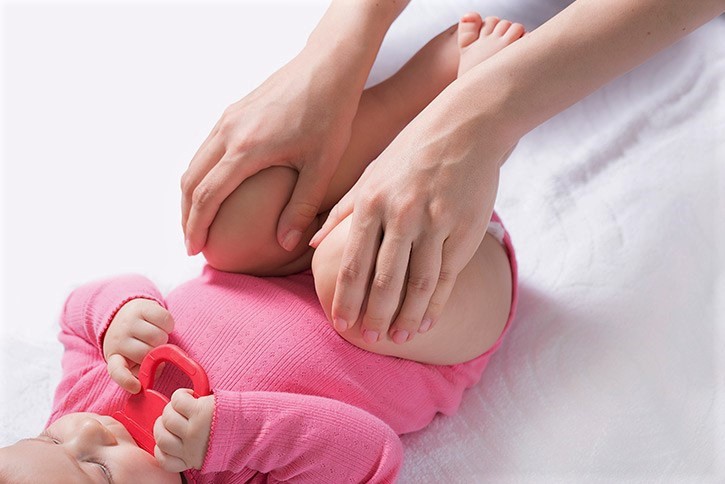
How it is recognised in children
For children, manoeuvres are also used, which, unfortunately, also lose sensitivity as they get older and their joints develop.
The doctor goes and stretches the hip, which, if affected by dysplasia, produces characteristic noises.
The most well-known manoeuvres are:
- Ortolani manoeuvre (named after the paediatrician who invented it): when, as a result of certain movements, the head of a femur that is not fully positioned in the acetabulum is repositioned inside it, it emits a snap;
- Barlow’s manoeuvre (by orthopaedist T.G. Barlow): when, as a result of the doctor’s movements, the head of a femur that is located in the acetabulum, but is not lodged in it correctly, protrudes from it, resulting in a snap.
Diagnostic imaging in cases of hip dysplasia
The objective examination and manoeuvres depend very much on the sensitivity and ability of the doctor; therefore, the key tests for detecting hip dysplasia are those of diagnostic imaging and specifically
- ultrasound scan: this is the standard examination for the diagnosis of hip dysplasia in infants up to 3-4 months of age. It is recommended as a screening within the first 3 months; not too early as there can also be physiological delays in the development of the child’s joint. If, however, heredity and risk factors for luxation are present, it is recommended that it be performed within the first 6-8 weeks of life.
- X-ray: if indicated by the paediatrician or orthopaedic specialist, it is performed in adults and children over 3-4 months of age, as this is the period in which the ossification of the joint can be detected by X-ray.
- CT scan: it is mainly performed during therapy also to plan and evaluate the outcomes of prosthetic implants.
The symptoms of hip dysplasia
The symptoms of hip dysplasia in childhood are often very few, such as
- uneven leg length
- asymmetry in the skin folds of the thighs;
- reduced mobility and flexibility in the lower limbs on one side of the body compared to the other.
In particularly severe or degenerate cases, this may also be characterised by:
- joint pain;
- lameness;
- inability or difficulty in performing certain movements such as crossing the legs;
- instability.
The causes of DCA are not yet known, but factors related to:
- heredity, particularly as regards the female sex, which is more exposed, and the left side of the body or both sides;
- birth in a breech position (with the head placed upwards, instead of towards the mouth of the uterus);
- coexistence of other malformations such as club foot, flat foot etc.
Consequences of hip dysplasia
It is important for hip dysplasia to be diagnosed as early as possible to allow for correction in the developmental and osteo-articular phases of the child.
If the condition is not treated in these early stages, in fact:
- if it is of a moderate degree, it can lead to early arthrosis, even in the young adult, so-called coxarthrosis. This pathology can, however, also affect an early corrected joint that, although improved, has failed to achieve a normal appearance and development ;
- if severe, it can soon lead to dislocation resulting in shortening of the limb, joint limitation and lameness.
How hip dysplasia is treated
Once a diagnosis has been made, treatment for hip dysplasia varies depending on the severity of the condition and the age of the subject, following a conservative approach wherever possible.
Alternatively, in the most severe cases, surgical therapy is carried out.
Conservative therapy in children
In children up to 6 months with mild to medium cases, in most cases, retractors are prescribed, i.e., braces of different types and characteristics (Pavlik retractor, Milgram retractor, Tübingen retractor, etc.) that, as the word itself indicates, spread and flex the child’s legs, immobilising them in a position that allows the femoral head to retract into the acetabulum, taking advantage of growth stimuli to improve the development and conformation of the joint.
Conservative therapy in adults

With regard to adults, in cases where the severity of the pathology allows it, joint infiltrations of autologous substances, i.e. the patient’s own substances, can be performed, which have an anti-inflammatory and regenerative action.
The most common of these substances are:
- PRP (Platelet Rich Plasma): a small amount of blood is taken from the patient which, cleansed of impurities, is rich in platelets;
- components of adipose tissue which, taken by liposuction (in the operating theatre) and appropriately purified, are rich in stem cells.
Surgical therapy in children
Reductions and osteotomies are performed in children
- over the age of 6 months
- with severe DCA for which retractor therapy has not worked or is unsuitable;
- in the presence of a dislocation that cannot be manually retracted.
The only solution is surgery with an operation performed in the operating theatre, which can be one:
- Closed (or non-blood) reduction: the paediatric orthopaedist manually centres and moves the femur inside the acetabulum without making any major cuts;
- Open (or cruciate) reduction: used in the most severe cases. A more significant cut is made to allow the surgeon to position the femoral head in the acetabulum correctly or as angled as possible. An open reduction may also be accompanied by osteotomies, which are procedures to reorganise the femoral-acetabular area by cutting bones to reposition them and correct structural deformities.
After surgery, the child is usually fitted with a cast to keep the hip in the correct position during healing.
The surgical procedure may or may not also be preceded by gradual traction of the hips.
Surgical therapy in adults
It is possible to proceed through:
- hip arthroscopy;
- hip replacement.
Hip arthroscopy
Hip arthroscopy is a minimally invasive technique that uses very small incisions to insert an arthroscope into the area to examine the joint from the inside, and to operate on it.
The anatomical space of the hip is very limited, so the lower limb is placed in traction so that there is enough of an opening to pass the arthroscope and instruments for corrective procedures.
Hip prosthesis
As far as adults are concerned, recourse to a hip arthroplasty is necessary to restore the correct function of the joint in cases that are unsuitable or unresponsive to conservative therapy of:
- undiagnosed and uncorrected DEA in childhood with pain and mobile difficulties or even wear/damage of parts of the joint;
- partially corrected joint, also in childhood, which has suffered arthrosis;
- evolution of the pathology into dislocation of the hip, which cannot be manually retracted.
From an anterior or antero-lateral access, which almost completely eliminates the risk of dislocation, an incision of approximately 15-20 cm is made, through which the metal prosthesis (generally titanium) is passed, without touching the musculature, which can
- cover only the head of the femur, which is then otherwise preserved;
- replace the entire bone/cartilage of the femur and the housing acebular cavity which, therefore, are totally removed.
The prosthesis can be cemented to the natural bone, but in Italy a biological approach is preferred whereby the body adapts naturally to the new inserted structure.
Hip prostheses allow an optimal quality of life and can last even more than 20 years.
The patient may subsequently need physiotherapy to regain proprioception, i.e. sensitivity in space, but can generally move as early as the day of surgery, with a considerable reduction in pain compared to pre-operatively.
Read Also
Emergency Live Even More…Live: Download The New Free App Of Your Newspaper For IOS And Android
Hip Osteoarthritis: What Is Coxarthrosis
Why It Comes And How To Relieve Hip Pain
Hip Arthritis In The Young: Cartilage Degeneration Of The Coxofemoral Joint
Visualizing Pain: Injuries From Whiplash Made Visible With New Scanning Approach
Coxalgia: What Is It And What Is The Surgery To Resolve Hip Pain?
Unicompartmental Prosthesis: The Answer To Gonarthrosis
Shoulder Instability And Dislocation: Symptoms And Treatment