
Impetigo, how to recognise it and how to treat it
Impetigo belongs to the large group of pyoderma, pustular infections of the skin caused by pyogenic (i.e. pus-producing) germs
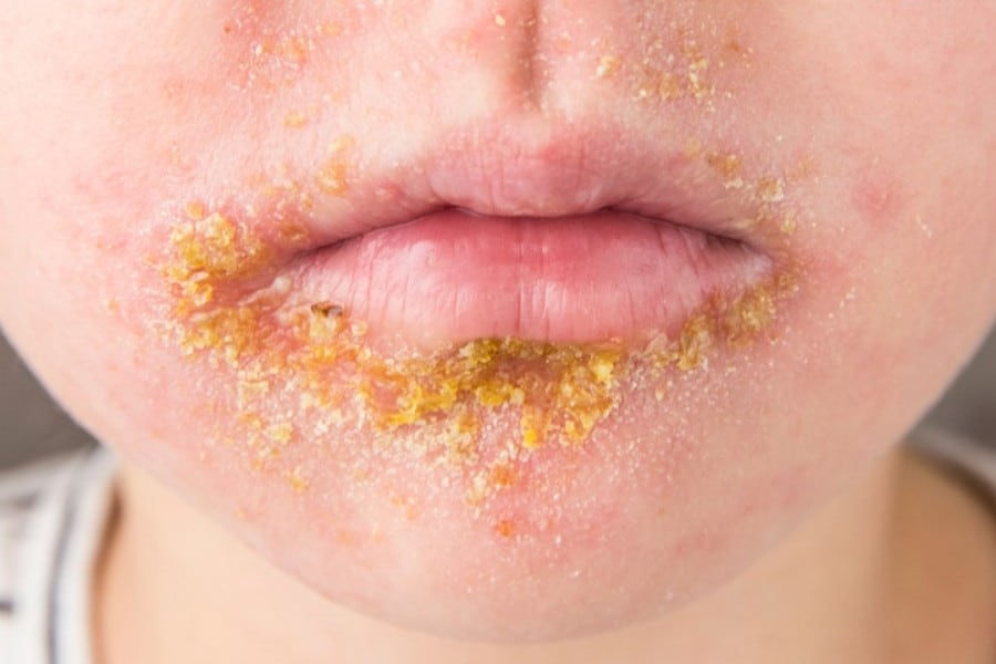
It is a disease characterised by vesicles and pustules that arise on healthy skin, are inoculable and autoinoculable (i.e. they are transmitted by contact from sick to healthy and multiply in the same patient by touching), heal forming a yellowish crust that, once fallen off, leaves no scars.
Causes of infection
The two microbes responsible for impetigo are staphylococcus and streptococcus, both of which are often present at the same site, so it is difficult to determine which is the primary germ and which is the secondary colonising germ.
The distinction is important as the treatment of the two bacteria is different.
Clinical forms of impetigo
Traditionally, impetigo is distinguished into two clinical forms that differ in both epidemiology and clinical manifestations.
Tilbury-Fox impetigo, also known as impetigo vulgaris or streptococcal impetigo
It is caused by streptococcus and generally affects children aged 5 to 7 years, with a peak incidence during August and September and a second peak in early spring, regardless of gender.
Among adults, it prevails instead in the male sex, with possible epidemics in community places (barracks, seminaries): in these cases the contagiousness is very high, and not necessarily linked to poor hygiene (which in any case predisposes to contagion).
Bockart’s impetigo, also called staphylococcal impetigo, acute ostium-follicular impetigo or bullous impetigo
This is the most frequent form, generally caused by the settlement of staphylococcus, without association with other bacteria, inside the skin’s follicular ostium.
It affects all ages, with maximum incidence during the summer, and normally manifests itself as an isolated case, although small epidemics may arise due to living in the family or in crowded communities (nurseries, schools).
It is not linked to the hygienic conditions of the skin but to the presence of minimal excoriations or scratching lesions, which favour the establishment of the bacterium.
Impetigo: What are the symptoms?
Tilbury-Fox impetigo usually begins with the formation of a single vesicle, 4-5 mm in diameter, filled with a clear serous fluid that soon becomes cloudy and purulent.
The dome of the vesicle sags and, being very thin, is easily ruptured, allowing the serous or purulent contents to leak out, forming a yellow crust on a reddened area of skin.
If the size of the vesicle is conspicuous we speak of bullous impetigo, and next to the initial vesicle/bubble subsequent lesions may easily arise, which tend to converge, giving rise to extensive crusts with polycyclic margins.
The disease favours the face, with rapid dissemination of the elements by autogenous grafting.
The evolution of each individual element lasts 4-8 days, leaving a reddened patch at the fall of the scab that disappears completely with time.
This course is typical of the disease treated with antibiotics: in the absence of treatment, impetigo may last for weeks or months, with recurrent fever flares and regional lymphatic resentment.
Bockart’s impetigo is characterised by vesicular elements that from their onset are filled with a creamy, dense, greenish-yellow content.

In contrast to the vesicles of the previous form, these are very resistant and unlikely to rupture, forming a small dome that is often situated at the level of a follicular ostium and, in this case, is centred by a hair.
They are always surrounded by a reddened halo.
The evolution is like that of the previous form, but the infection has no predilection of site or age.
Any agent that alters the integrity of the skin (scratching lesions, excoriations, chemical irritants) is a favourable factor.
How to diagnose impetigo
It is not difficult: the presence of vesicular and crusty elements, with dissemination by self-inoculation, at an asymmetrical site easily points towards the diagnosis.
The content of the vesicles and their morphology also suggest which bacterium is involved: fragile vesicles, with serous content in the streptococcal forms; resistant vesicles, with purulent greenish-yellow content in the staphylococcal forms.
Therapy for impetigo
Effective local therapy on cleansed skin is sufficient to eradicate impetigo: aseptically open the vesicles and pustules that are still closed, remove the scabs and dress with antiseptic or antibiotic ointments.
For bacteriological reasons, however, oral antibiotics should be preferred to the ‘old’ penicillin or erythromycin, effective on both streptococcus and staphylococcus.
Read Also:
Emergency Live Even More…Live: Download The New Free App Of Your Newspaper For IOS And Android
Herpes Zoster, A Virus Not To Be Underestimated
Shingles, The Painful Return Of The Chickenpox Virus
What Is Impetigo In Adults And Children And How To Treat It
Ramsay Hunt Syndrome: Symptoms, Treatment And Prevention
What Is Impetigo In Adults And Children And How To Treat It
Shingles: Symptoms, Causes And How To Ease The Pain
Breastfeeding Women And Vaccine, The Pediatrician Assures: “It Is Effective And Recommended”
Healing Wounds And Perfusion Oximeter, New Skin-Like Sensor Can Map Blood-Oxygen Levels
SkinNeutrAll®: Checkmate For Skin-Damaging And Flammable Substances