
Paediatric Intubation: Achieving a Good Outcome
Paediatric intubation is a procedure that must be fully understood: prematurity and immature development of the pulmonary system, severe illness, trauma, overdose, aspiration, and other cardiopulmonary conditions call for protection of the airway and establishment of a safe and secure means to ventilate
Infants and children undergo changes in the anatomy as they grow, with the differences most pronounced under 2 years of age
Between 2 and 8 years old the airway is in transition, and by age 8 the anatomy is comparable to a small adult.1
Children have several distinctions in the airway anatomy including a larger tongue, a larger and floppy epiglottis, a narrow cricoid ring, and a higher and more anterior glottic opening.
At infancy, the glottis is located at C1, moving downward to C3-C5 around age 7, and finally reaching C4-C6.1
The trachea is shorter, more narrow, and more compliant.2
From birth to about 2 years old there is less control of respiration, inefficient use of the muscles of ventilation, different airway and lung mechanics, and a higher basal metabolic rate (increasing the risk of rapid desaturation during intubation).
These factors result in higher risk of cardiorespiratory failure, which is even more pronounced during an intubation procedure in neonatal and pediatric patients.
The total lung capacity (TLC) and functional residual capacity (FRC) is lower, and the thorax is more compliant while the lungs are less compliant.
These issues bring about increased tendency for airtrapping and early closure of the terminal airways.2
Preparing for Paediatric Intubation
Training for neonatal/pediatric intubation can be done using simulation to allow for a controlled, safe setting, provide time for the team to improve communication, practice using different aids to intubation, and work through different scenarios to prepare for changing conditions or special circumstances.
Having an intubation checklist has proven to be very helpful to be sure all the equipment is available and functioning, appropriate medications and respective dosages have been reviewed and prepared, and the intubation team is primed and ready for the procedure.
Sample checklists are readily available, and if needed, the team can customize these to fit the particulars of their institution.2-3
Assessment of the patient prior to intubation is needed to evaluate the anatomy and check on the hemodynamic and pulmonary status.
Due to the anatomic differences mentioned above, a shoulder roll or folded towel may be useful to position the head and neck so that the vocal cords and glottis can be visualized.
Historically, uncuffed ETT have been most commonly used in neonatal patients but use of cuffed tubes are increasing as studies have shown that they may be a better choice.4
Orotracheal intubation is also the most used route but nasotracheal intubation is an available option and may be a consideration as more studies are being published showing favorable outcomes.5
ETT sizes for neonates are often determined by patient weight (2.5 mm tube if <1,000 grams, 3.0 tube if 1,000-2,000 grams, 3.5 mm tube if 2,000-3,000 grams, 3.5 or 4.0 mm tube if >3,000 grams).3
Selecting the right size ETT has been a challenge, with several proposed size prediction models available.
An article published in 2022 compared four methods of estimating the correct size tube for pediatric patients and concluded that ultrasonic evaluation of the cricoid cartilage and trachea was the best approach, and using the diameter of the little finger to be the best of the conventional approaches.6
CHILD HEALTH: LEARN MORE ABOUT MEDICHILD BY VISITING THE BOOTH AT EMERGENCY EXPO
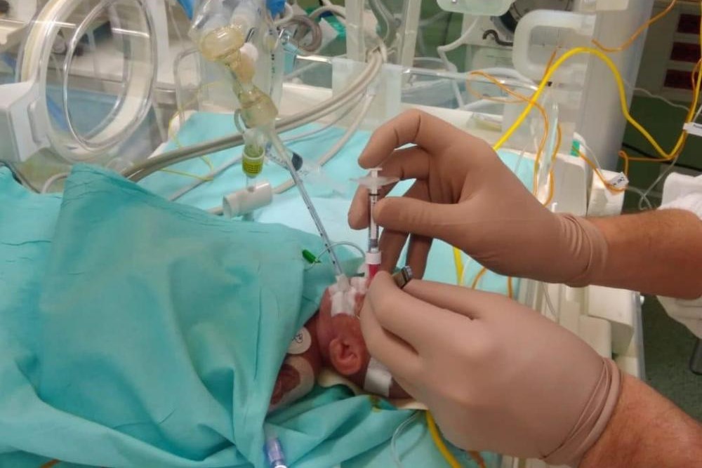
The Paediatric Intubation Procedure
To intubate, the patient should be properly positioned and preoxygenated.
If possible, medications (sedative and paralytic) may be useful to facilitate intubation.
Oxygen and bag-mask ventilation should be continued throughout the procedure to avoid desaturation and clear exhaled CO2.
Sedation is often accomplished with Fentanyl (4 mcg/kg) followed by saline flush given first, then consider giving Atropine (20 mcg/kg) followed by saline flush to reduce issues with reflex bradycardia and dry secretions.
Suxamethonium (2 mg/kg) followed by saline flush is given to paralyze, and the intubation should follow immediately.
The procedure should stop if the intubation is not completed within 30 seconds from the time of laryngoscope insertion, if bradycardia <70 beats/minute occurs at any time, or if oxygen saturation drops <70%.
Use bag-mask ventilation with oxygen to regain a good baseline before restarting the procedure.3 A bougie catheter may be used to establish the path through the glottis and allow for the ETT to pass over the catheter into the lower airway.
Video laryngoscopy may be helpful if the glottis cannot be visualized.6
Correct position in the airway is challenging in neonates and younger pediatric patients due to the short length of the trachea.
Prediction models have been used to give guidance for ETT depth but there have been several studies that show problems with the many of the guidelines. Work is continuing to try and improve these models.2,8
Confirmation of ETT placement includes physical examination, auscultation of the chest and epigastrum, capnography, chest radiography, use of light/sound transmitting devices, and ultrasound.2
Movement of the head (by neck flexation or neck extension or turning the head from side to side) moves the ETT up or down in the trachea.
This may malposition the ETT so extra care must be taken to insure the tube is at the correct spot and is not malpositioned when procedures or moving the patient (ie transport, holding, repositioning in the bed) occur.2
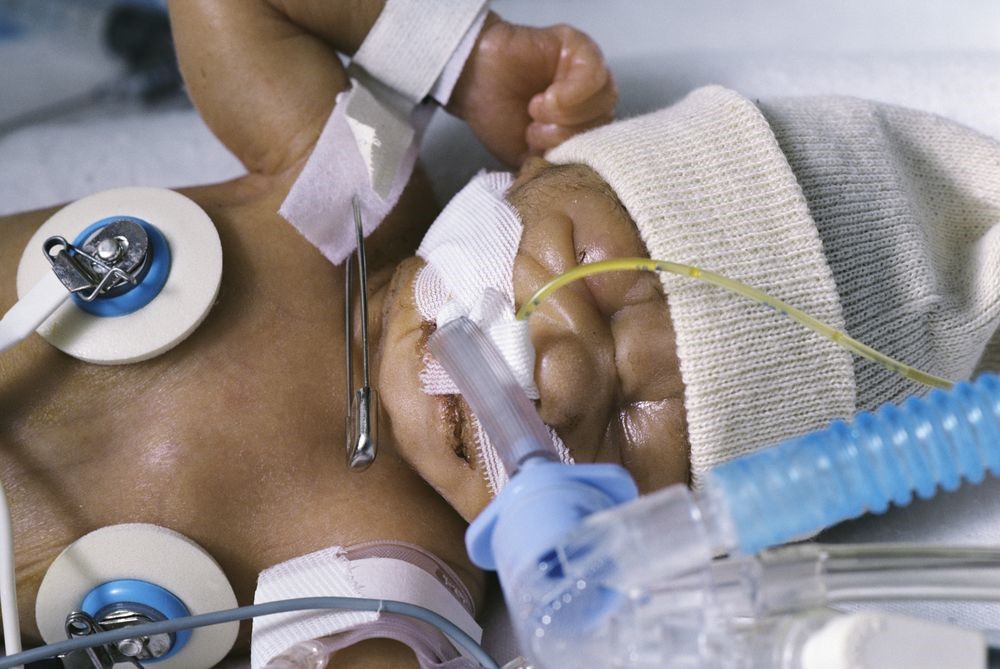
Securing the Airway
Once an ETT has been inserted and checked for proper position, it must be secured. ETT can be secured by taping or by a tube-holding device.
With either approach, extra care must be taken to avoid skin irritation, skin breakdown and pressure sores.
If using tape, use of “pre-taping” with a transparent dressing, such as 3M’s Tegaderm, Convatec’s Duoderm OpSite, or 3M’s No Sting barrier film, helps increase tape adhesion and protect the skin.
Several tapes are available to use including products such as Beiersdorf Inc’s Leukoplast or Elastoplast, or Kendall Healthcare Products’ WetPruf.
It is recommended that benzoin compounds not be used to aid in tape adherence to the skin for securing infant ETTs as damage to the skin may occur when removing the tape.9
Using tape to secure the ETT is difficult in neonates and pediatric patients due to the large amount of oral secretions
Many sites are using ETT holding devices that work effectively in securing the airway. There are a number of devices available on the market, including the AnchorFast ET Tube Holder, Cooper Surgical Neo-fit, Smiths Medical Portex ETTube Holder, Neotech Neo Bar, Ambu ETT Holder, and the Laerdal Thomas Tube Holder.9
Paediatric intubation, conclusion
Intubation in neonatal and pediatric patients is intense and can be stressful.
It requires skill, knowledge, practice, and the right team, right equipment, good communication, and good timing to be successful.
These patients cannot be cared for as “little adults” but call for special care due to their size and fragile nature.

There is less leeway, less wiggle room for having everything correct; placing the right-sized tube in the right position is more difficult.
References
- From the Anesthesia Key website: Intubation of the Pediatric Patient | Anesthesia Key (aneskey.com).
- Volsko TA. Kittredge Lecture: Airway Safety in Neonatal and Pediatrics. Respiratory Care. 2022 Jun 1;67(6):756-68.
- Abdelhadi AA, et al. Non-Emergent Endotracheal Intubation of the Newborn: Practical Management. 2022 Mar.
- Chen L, et al. Cuffed versus uncuffed endotracheal tubes in pediatrics: a meta-analysis. Open Medicine. 2018 Jan 1;13(1):366-73.
- Christian CE, et al. Use and outcomes of nasotracheal intubation among patients requiring mechanical ventilation across US PICUs. Ped Crit Care Med. 2020 Mar 11;21(7):620-4.
- Putra SR. Accuracy Comparison between Four Methods of Endotracheal Tube Diameter Estimation for Pediatric Patients: An Observational, Cross-sectional Study.
- Zhou M, et al. Video laryngoscopy improves the success of neonatal tracheal intubation for novices but not for experienced medical staff. Frontiers in Pediatrics. 2020 Aug 6;8:445.
- Volsko TA, et al. Development and internal validation of an equation using anthropometric measures to predict correct endotracheal tube insertion depth. Can J Resp Ther. 2022;58:9.
- Andrews D, et al. Securing paediatric endotracheal tubes: Tape it like you mean it! Australasian Emerg Nurs J. 2007 Mar 1;10(1):30-3.
- Atkins DL, et al. 2022 Interim Guidance to Health Care Providers for Basic and ACLS in Adults, Children, and Neonates With Suspected or Confirmed COVID-19. Circulation: Cardiovascular Quality and Outcomes. 2022 Apr;15(4):e008900.
Read Also
Emergency Live Even More…Live: Download The New Free App Of Your Newspaper For IOS And Android
How Common Is Brain Damage At Birth?
Head Trauma And Brain Injuries In Childhood: A General Overview
Neonatal/Pediatric Endotracheal Suctioning: General Characteristics Of The Procedure
Step-By-Step Guide To Intubation
Choking With Obstruction From Food, Liquids, Saliva In Children And Adults: What To Do?
Ventilator Management: Ventilating The Patient
Neonatal/Pediatric Endotracheal Suctioning: General Characteristics Of The Procedure
Three Everyday Practices To Keep Your Ventilator Patients Safe
Ambulance: What Is An Emergency Aspirator And When Should It Be Used?
The Purpose Of Suctioning Patients During Sedation
Supplemental Oxygen: Cylinders And Ventilation Supports In The USA
Basic Airway Assessment: An Overview
Respiratory Distress: What Are The Signs Of Respiratory Distress In Newborns?
EDU: Directional Tip Suction Catheter
Suction Unit For Emergency Care, The Solution In A Nutshell: Spencer JET
Airway Management After A Road Accident: An Overview
Tracheal Intubation: When, How And Why To Create An Artificial Airway For The Patient
What Is Transient Tachypnoea Of The Newborn, Or Neonatal Wet Lung Syndrome?
Asphyxia: Symptoms, Treatment And How Soon You Die
Choking (Suffocation Or Asphyxia): Definition, Causes, Symptoms, Death
First Aid For Scalding: How To Treat Hot Water Burn Injury
Step-By-Step Guide To Intubation
Electrical Burn: First Aid Treatment And Prevention Tips
Emergency Interventions: The 4 Stages Preceding Death By Drowning
Drowning Resuscitation For Surfers
ERC 2018 – Nefeli Saves Lives In Greece
First Aid In Drowning Children, New Intervention Modality Suggestion
Water Rescue Dogs: How Are They Trained?
Drowning Prevention And Water Rescue: The Rip Current
Water Rescue: Drowning First Aid, Diving Injuries
RLSS UK Deploys Innovative Technologies And The Use Of Drones To Support Water Rescues / VIDEO
Summer And High Temperatures: Dehydration In Paramedics And First Responders
First Aid: Initial And Hospital Treatment Of Drowning Victims
First Aid For Dehydration: Knowing How To Respond To A Situation Not Necessarily Related To The Heat
Children At Risk Of Heat-Related Illnesses In Hot Weather: Here’s What To Do
Summer Heat And Thrombosis: Risks And Prevention
Dry And Secondary Drowning: Meaning, Symptoms And Prevention
Drowning In Salt Water Or Swimming Pool: Treatment And First Aid
Water Rescue: Drone Saves 14-Year-Old Boy From Drowning In Valencia, Spain
Death By Suffocation: Signs, Symptoms, Stages And Timing