
Spine and lower limbs: what is EOS and when is it used?
EOS is a newly developed X-ray device that allows the spine and lower limbs to be studied in the upright (loaded) position
It makes use of ionising radiation to obtain digital images apparently similar to those of a normal X-ray machine, but with a significantly reduced exposure dose, and susceptible to three-dimensional reconstructions.
What is EOS used for?
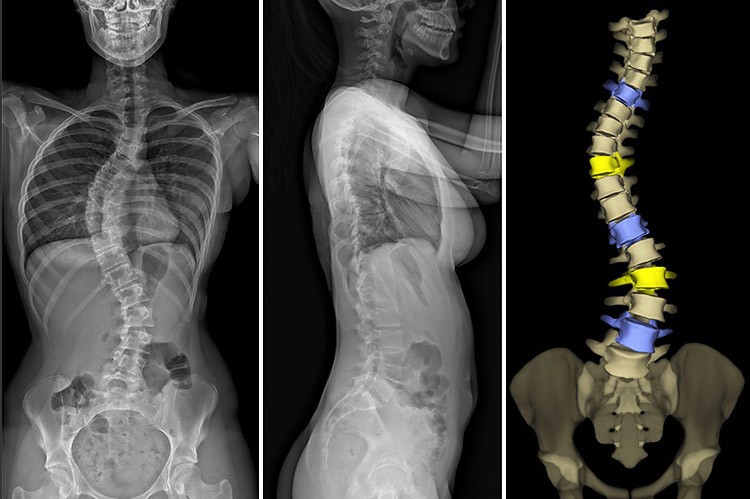
EOS can provide images of several anatomical districts but was created to study, together or separately, the spine, pelvic girdle and lower limbs, “in load” with the patient standing or sitting, on a single image obtained in a few seconds of exposure.
The images provided by EOS, although apparently similar, are diagnostic images that cannot be obtained with conventional radiology, because they are not subjected to magnification and photographic distortion; they also allow an evaluation of the entire axial skeleton thanks to a field of view of 175 cm on a 1:1 scale, with a significantly lower dose level (8 to 10 times) than conventional radiological techniques and not even comparable to an equivalent test possibly performed with CT (dose delivered 800/900 times lower).
Who can perform the EOS test?
Particularly indicated for the study of spinal pathologies in children and the elderly for the evaluation of posture parameters, it offers orthopaedic surgeons and neurosurgeons valuable information for targeted and preventive intervention.
The pathologies that benefit more from the use of EOS compared to traditional techniques (scoliosis, spinal and hip defects, congenital or acquired skeletal dysmorphia) mainly affect young patients or children who need frequent repeated checks over time.
Hence the need for special attention to be paid to dosimetric aspects.
The patient must be able to remain upright during the test.
Is EOS painful or dangerous?
EOS is not a painful test and, although it uses radiation, it is to date the least dangerous tool in use for obtaining diagnostic images of the spine due to the significantly lower dose compared to traditional reference methods (8/10 times).
How does it work?
Performing a test with Eos presents, on a practical level, some differences compared to traditional radiology. First of all, the machine looks different.
In order to obtain the images, the patient has to enter a sort of cabin, where he or she stands, still, for about twenty seconds while the scan is taken.
The other ‘rules’ customary in radiology remain valid, such as the need to leave any metal objects outside the machine or the need to wear protection for the most sensitive areas to radiation, such as the genitals, despite the considerably lower doses to which one is exposed.
The images are completely digital (no X-ray cassettes or films are used) and are managed by dedicated software that generates 3D reconstructions and/or volumetric renderings to study pathologies (of the spine and limbs) linked to problems of dynamics and posture (basic and loaded).
The EOS technology is linked to the name of a Nobel Prize winner in physics, Georges Charpak, who developed the special detectors that enable the high signal-to-noise ratio images provided by the machine to be obtained at minimum doses.
Read Also
Emergency Live Even More…Live: Download The New Free App Of Your Newspaper For IOS And Android
Radiography: The Role Of X-Ray In Bone And Soft Tissue Diagnosis
What Is Hand Radiography (Hand X-Ray)?

Bone Scintigraphy: How It Is Performed
Radiography: What It Is And What It Consists Of
What Is Hand Radiography (Hand X-Ray)?
Intraosseous Access, A Life-Saving Technique In Emergency Shock Management
Electromyography (EMG), What It Assesses And When It Is Done
Dislocation Of The Shoulder: How To Reduce It? An Overview Of The Main Techniques
Fusion Prostate Biopsy: How The Examination Is Performed
CT (Computed Axial Tomography): What It Is Used For
What Is An ECG And When To Do An Electrocardiogram
Positron Emission Tomography (PET): What It Is, How It Works And What It Is Used For
Single Photon Emission Computed Tomography (SPECT): What It Is And When To Perform It
Instrumental Examinations: What Is The Colour Doppler Echocardiogram?
Coronarography, What Is This Examination?
CT, MRI And PET Scans: What Are They For?
MRI, Magnetic Resonance Imaging Of The Heart: What Is It And Why Is It Important?
Urethrocistoscopy: What It Is And How Transurethral Cystoscopy Is Performed
What Is Echocolordoppler Of The Supra-Aortic Trunks (Carotids)?
Surgery: Neuronavigation And Monitoring Of Brain Function
Robotic Surgery: Benefits And Risks
Refractive Surgery: What Is It For, How Is It Performed And What To Do?