
Radiography: the role of X-ray in bone and soft tissue diagnosis
An X-ray is an imaging study that takes pictures of bones and soft tissues. X-rays use safe amounts of radiation to create these pictures
The images help healthcare providers diagnose a wide range of conditions and plan treatments.
Usually, providers use X-rays to evaluate broken bones, dislocated joints and other bone injuries.
What is an X-ray study?
An X-ray study (also called a radiograph) is a type of medical imaging (radiology) that creates pictures of your bones and soft tissues, such as organs.
X-rays use safe amounts of radiation to make these pictures.
The images help your provider to diagnose conditions and plan treatments.
Most often, providers use X-rays to look for fractures (broken bones).
But X-ray images can help providers diagnose a wide range of injuries, disorders and diseases.
X-rays are a safe and effective way for providers to evaluate your health.
Who might need an X-ray?
People of all ages, including babies, can get an X-ray.
If there’s a chance you might be pregnant, tell your provider before getting an X-ray.
Radiation from an X-ray can harm your fetus.
Your provider may order an X-ray to:
- Check for a broken bone (fracture).
- Identify the cause of symptoms, such as pain and swelling.
- Look for foreign objects in your body.
- Look for structural problems in your bones, joints or soft tissues.
- Plan and evaluate treatments.
- Provide routine screenings for cancer and other diseases.
What are the types of X-ray studies?
Several types of X-rays take pictures of different areas inside your body.
Some X-rays use contrast material (also known as dye) to make the images clearer.
Some of the most common types of X-rays include:
- Abdominal X-ray: This X-ray shows images of your kidneys, stomach, liver and bladder. It helps providers diagnose conditions like kidney stones and bladder stones. There are some special kinds of abdominal X-rays such as a barium enema that use special dyes (called contrast) to evaluate parts of the digestive system.
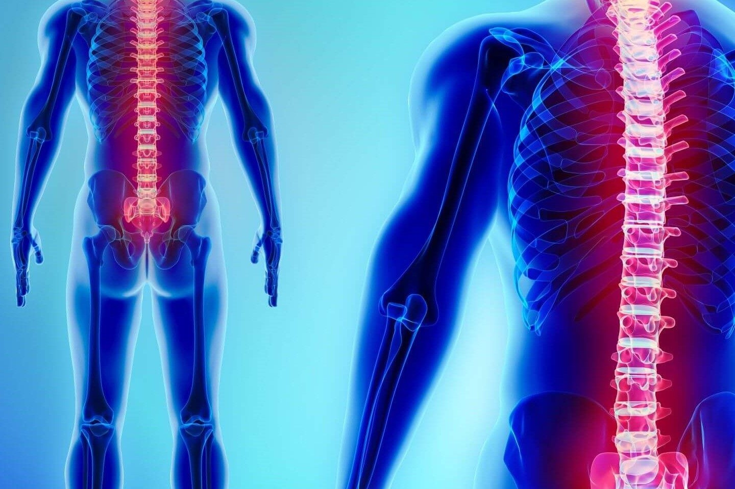
- Bone X-ray: Your provider uses a bone X-ray study to see broken bones (fractures), dislocated joints and arthritis. Images from bone X-rays can also show signs of bone cancer or infection. A spine X-ray looks at the bones and tissues in the spine.
- Chest X-ray: This test looks for abnormalities in the heart, lungs and bones in the chest like pneumonia.
- Dental X-ray: Regular dental X-rays allow your provider to evaluate your teeth and gums, look for infection and check for cavities.
- Fluoroscopy: A fluoroscopy shows moving images of organs and soft tissues (such as your intestines). Your provider views your organs in motion on a screen (kind of like an X-ray movie). GI X-ray exams often use fluoroscopy.
- CT scan (computed tomography): A radiology study that uses X-rays and a computer to create cross-section images of bones, organs and tissues. This is a donut-shaped machine that you slide through as it takes images.
- Mammogram: Providers use mammograms to take X-ray pictures of breast tissue, evaluate breast lumps and diagnose breast cancer.
What is an X-ray with contrast material?
Some X-rays use contrast material (also called contrast agent or dye).
The contrast material comes as a liquid, powder or pill.
Your provider gives you the contrast material before the X-ray.
Depending on the type of X-ray, you may receive the contrast material:
- Orally (by mouth).
- Through an injection like from an intravenous (IV) shot.
- By inserting it into your rectum (enema).
When your provider gives you the dye through an IV injection, you may feel flushed or warm for a little while. Some people experience a metallic taste in their mouth. These side effects go away in a few minutes.
The contrast agent changes the way soft tissues and other structures appear on an X-ray study so your provider can see them in more detail.
How does an X-ray study work?
An X-ray sends beams of radiation through your body.
Radiation beams are invisible, and you can’t feel them.
The beams pass through your body and create an image on an X-ray detector nearby.
As the beams go through your body, bones, soft tissues and other structures absorb radiation in different ways.
Solid or dense objects (such as bones) absorb radiation easily, so they appear bright white on the image.
Soft tissues (such as organs) don’t absorb radiation as easily, so they appear in shades of gray on the X-ray.
How do I prepare for an X-ray?
Tell your healthcare provider about your health history, allergies and any medications you’re taking.
If you’re pregnant, think you might be pregnant or are breastfeeding (chestfeeding), tell your provider before getting an X-ray.
You usually don’t need to do anything to prepare for a bone X-ray.
For other types of X-ray, your provider may ask you to:
- Avoid using lotions, creams or perfume.
- Remove metal objects like jewelry, hairpins or hearing aids.
- Stop eating or drinking several hours beforehand (for GI X-rays).
- Wear comfortable clothing or change into a gown before the X-ray.
What should I expect during an X-ray?
Depending on the type of X-ray, your provider will ask you to sit, stand or lie down on a table.
During the X-ray, your provider may move your body or limbs in different positions and ask you to hold still.
You may need to hold your breath for a few seconds so the images aren’t blurry.
Sometimes children can’t stay still long enough to produce clear images.
Your child’s provider may recommend using a restraint during an X-ray.

The restraint (or immobilizer) helps your child stay still and reduces the need for retakes.
The restraints don’t hurt and won’t harm your child.
What should I expect after an X-ray?
If you received contrast dye before your X-ray, you should drink plenty of water to flush the contrast material from your body.
Some people have side effects from contrast dye, which may include:
- Nausea or vomiting.
- Stomach cramps or diarrhea.
- Headaches.
- Rarely, allergic reactions to contrast material can occur. People who have allergies or asthma are more likely to have an allergic reaction to contrast dye. Talk to your provider about your risk of a reaction, and call your provider right away if you have unusual symptoms.
What are the risks of an X-ray?
Although X-rays use radiation (which can cause cancer and other health problems), there is a low risk of overexposure to radiation during an X-ray.
Some X-rays use higher doses of radiation than others.
Generally, X-rays are safe and effective for people of all ages.
Radiation from an X-ray can harm your fetus.
If you’re pregnant, your provider may choose another imaging study, such as MRI or ultrasound.
When should I know the results of my X-ray?
Results from a bone X-ray are usually ready right away.
Your provider may share your results with you after the X-ray.
Results from other types of X-rays (such as a GI test) may take longer.
Talk to your provider about when you can expect results.
When should I call my doctor?
Allergic reactions to contrast material are rare.
Symptoms can appear up to a day or two after the X-ray.
If you received contrast material before your X-ray, call your provider if you have:
- Skin rash, hives or itching.
- Headaches.
- Nausea or vomiting.
- Trouble breathing or shortness of breath.
Read Also
Emergency Live Even More…Live: Download The New Free App Of Your Newspaper For IOS And Android
What Is Hand Radiography (Hand X-Ray)?
Bone Scintigraphy: How It Is Performed
Radiography: What It Is And What It Consists Of
What Is Hand Radiography (Hand X-Ray)?
Intraosseous Access, A Life-Saving Technique In Emergency Shock Management
Electromyography (EMG), What It Assesses And When It Is Done
Dislocation Of The Shoulder: How To Reduce It? An Overview Of The Main Techniques
Fusion Prostate Biopsy: How The Examination Is Performed
CT (Computed Axial Tomography): What It Is Used For
What Is An ECG And When To Do An Electrocardiogram
Positron Emission Tomography (PET): What It Is, How It Works And What It Is Used For
Single Photon Emission Computed Tomography (SPECT): What It Is And When To Perform It
Instrumental Examinations: What Is The Colour Doppler Echocardiogram?
Coronarography, What Is This Examination?
CT, MRI And PET Scans: What Are They For?
MRI, Magnetic Resonance Imaging Of The Heart: What Is It And Why Is It Important?
Urethrocistoscopy: What It Is And How Transurethral Cystoscopy Is Performed
What Is Echocolordoppler Of The Supra-Aortic Trunks (Carotids)?
Surgery: Neuronavigation And Monitoring Of Brain Function
Robotic Surgery: Benefits And Risks
Refractive Surgery: What Is It For, How Is It Performed And What To Do?