
Radiography: what it is and what it consists of
Radiography is a diagnostic technique that relies on the use of X-rays and the ‘braking’ effect due to the interaction between matter and radiation
Invented by Wilhelm Conrad Röntgen in 1895, traditional radiography was the first biomedical imaging technique and has undergone numerous developments and evolutions over the years, culminating in computerised radiography.
What does radiography consist of
The X-ray examination is performed by a doctor specialising in radiology or a radiology technician.
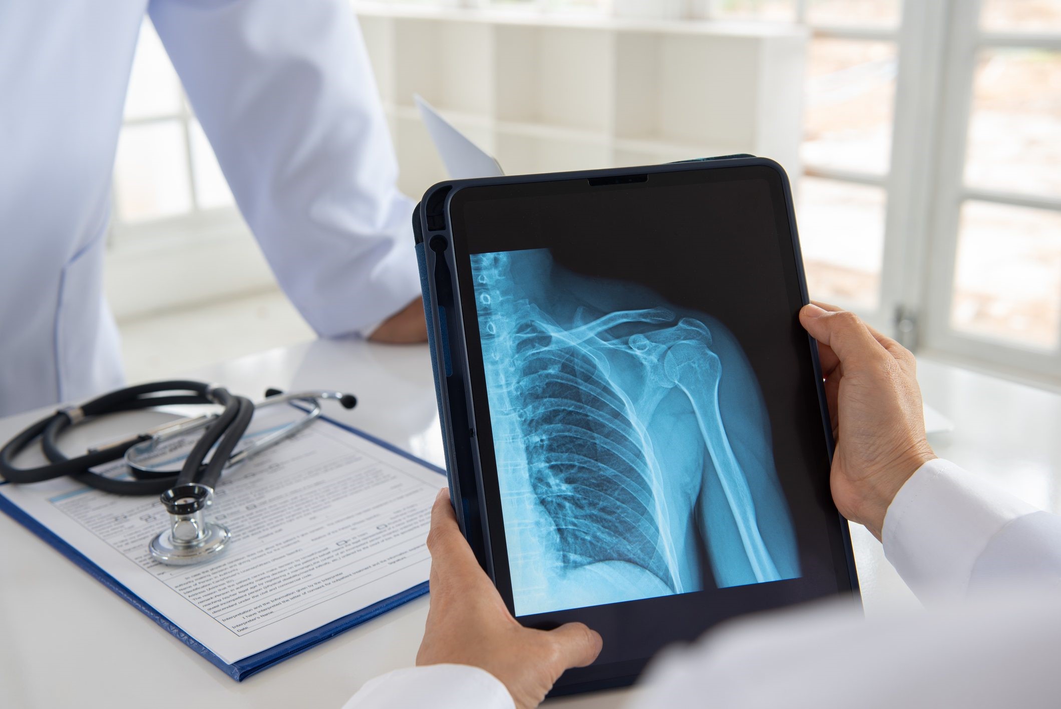
In traditional radiography, the part of the body to be analysed is brought into contact with the radiation-generating machine.
The X-ray beam passes through the patient’s body and impresses the X-ray film, on which an image is formed that enables the structures and tissues concerned to be distinguished: the X-ray image is determined because different body tissues absorb X-rays differently.
It is also possible to use radio-transparent or radio-opaque contrast media that emphasise the contours of the observed organ, making it easier to analyse.
Why and when to take a radiography
X-ray is a quick and easy examination, which is why it is usually used as the first diagnostic tool.
In bones, X-rays can detect fractures, lesions and tumours.
In the lungs, they can diagnose pneumonia or cancer.
In the case of a gunshot or foreign body injury, it may be able to detect the location of shrapnel or bullets.
For dentists, it is a basic resource for detecting cavities and the position of unbared teeth.
In bone densitometry, X-rays detect mineral deficiency in bones due to osteoporosis.
Useful to know about X-rays
X-rays can have a detrimental effect on certain anatomical areas, such as the ovaries and testicles, bone marrow and tissue in the process of formation.
This is why their use is now more limited and precautions are used, such as the use of specific lead shielding to protect the ovaries of women of childbearing age.
The health worker is also in an adjoining room during the X-ray.
Although the single dose of X-rays required for the examination is limited, over time the radiation would eventually accumulate and be harmful.
Read Also

Emergency Live Even More…Live: Download The New Free App Of Your Newspaper For IOS And Android
What Is Hand Radiography (Hand X-Ray)?
Bone Scintigraphy: How It Is Performed
Fusion Prostate Biopsy: How The Examination Is Performed
CT (Computed Axial Tomography): What It Is Used For
What Is An ECG And When To Do An Electrocardiogram
Positron Emission Tomography (PET): What It Is, How It Works And What It Is Used For
Single Photon Emission Computed Tomography (SPECT): What It Is And When To Perform It
Instrumental Examinations: What Is The Colour Doppler Echocardiogram?
Coronarography, What Is This Examination?
CT, MRI And PET Scans: What Are They For?
MRI, Magnetic Resonance Imaging Of The Heart: What Is It And Why Is It Important?
Urethrocistoscopy: What It Is And How Transurethral Cystoscopy Is Performed
What Is Echocolordoppler Of The Supra-Aortic Trunks (Carotids)?
Surgery: Neuronavigation And Monitoring Of Brain Function
Robotic Surgery: Benefits And Risks
Refractive Surgery: What Is It For, How Is It Performed And What To Do?