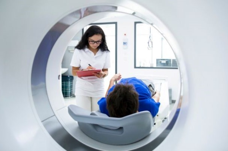
Adverse reactions to contrast medium: what are they and what incidence do they have?
Contrast medium is a substance used during radiological tests to improve the visibility of body structures by increasing the contrast between various organs and tissues
Incidence of side effects
Although they are considered safe, side effects to contrast media can sometimes occur.
The estimated incidence of radiological complications attributed to the administration of contrast media (MdC) is fortunately quite low, with values of less than 1% and a mortality rate of 1/60,000 tests.
Most reactions are idiosyncratic and unpredictable, but a careful anamnesis may make it possible to identify certain risk factors and allow an effective prophylaxis and immediate treatment plan to be set up from the very first symptoms, particularly in the case of an anaphylactic reaction, which can otherwise quickly lead to shock.
For this reason, treatment protocols inspired by the Guidelines, internationally recognised as valid for emergency-urgent conditions, must be followed in the diagnostic-therapeutic pathway.
Among the various contrast media, the most commonly used and most often responsible for adverse reactions are iodinated contrast media, which are mainly used in the angiological, urological and gynaecological fields.
Currently, where possible, so-called third-generation non-ionic contrast media are used, which are much more tolerable than ionic ones.
Reactions can occur immediately, but delayed reactions can also occur after an hour or sometimes up to a week.
What are the possible side effects to contrast media?
Adverse reactions to contrast medium are subdivided into:
- Chemotoxic (type A). They are so called because the toxicity of the compound is related to its chemical composition. These reactions are dependent on the dose and plasma concentration of the drug and are therefore potentially predictable.
- Anaphylactoids (type B). These are those in which the cause-effect relationship is most difficult to establish, are not dose-dependent and are by definition unpredictable (they may induce the release of histamine or other mediators usually active in allergic phenomena).
Depending on their severity, adverse reactions to contrast medium are subdivided into:
- mild: metallic taste, feeling of heat, nausea and vomiting, sweating, perioral dysesthesia, pain at the injection site, urticaria, migraine;
- moderate: persistence and increase in intensity of minor symptoms, dyspnoea, hypotension, chest pain
- severe: bronchospasm, anxiety, diarrhoea, paresthesias, localised and non-localised oedema, dyspnoea, cyanosis, marked hypotension, bradycardia, shock, pulmonary oedema, arrhythmias, convulsions, paralysis, coma, death.
Risk factors for adverse reactions to contrast medium
The following may be considered potentially at risk
- subjects with a known history of reaction to contrast media;
- asthmatic subjects and subjects with allergies who rely on continuous and periodic drug treatment;
- subjects with a latex allergy;
- very young or very old subjects, in which case the patient’s age is the potential risk factor;
- women.
Careful assessment of the patient’s clinical state appears essential: reduced renal and cardiovascular function are the real risk factors.
In light of the observations that the most severe reactions to contrast media may be sustained by an anaphylactic mechanism, some studies suggest that all patients with previous episodes of adverse reaction to contrast media should undergo a thorough allergological examination.
However, there are no tests that can predict the occurrence of side effects to contrast medium administration.
Can contrast medium side effects be prevented?
Studies on treatment with corticosteroids and antihistamines for patients at risk of an adverse reaction to contrast medium have shown conflicting results.
There is to date no randomised study in humans or animals that has proven the effectiveness of prophylaxis with antihistamines and/or corticosteroids in preventing reactions.
It was observed that premedication with corticosteroids and antihistamines reduced the incidence of mild adverse reactions, without changing the incidence of more severe reactions.
For patients who have shown severe adverse reactions, it is essential to administer ad hoc treatment within the first minute of the onset of symptoms.
The use of non-ionic hypo-osmolar contrast media and premedication, although carried out with different procedures and modalities, did not completely eliminate the likelihood of adverse reactions.
Contrast medium and renal failure
Contrast medium-induced nephropathy deserves a separate chapter.
Contrast medium can cause vasoconstriction of the renal tubular artery and alteration of glomerular haemodynamics.
In general, serum creatinine should be measured and glomerular filtration rate (GFR) calculated before administering contrast media in all patients:
- In adequately hydrated patients with normal renal function, acute renal failure is unlikely to occur.
- In patients with mild renal impairment, hydration before contrast medium administration usually prevents worsening of the renal picture.
- In patients with moderate to severe renal impairment, however, alternative instrumental investigations should be considered.
- In diabetic patients treated with metformin, discontinuation of the drug at least 12 hours before a contrastography test is recommended; this is because metformin has been associated with several cases of renal failure and lactic acidosis in patients exposed to contrast media.
Therefore, to reduce the risk of contrast media-induced nephropathy, it is important:
- Creatinine dosage and GFR calculation;
- Avoid repeated administration of high doses at short intervals;
- Hydrate the patient adequately intravenously if necessary;
- Use non-ionic contrast media with low osmolarity;
- Discontinue treatment with oral hypoglycaemic agents at least 12 hours before the MDC test;
- Avoid concomitant use of drugs that may cause renal vasoconstriction (e.g. NSAIDs).
In most cases of nephrotoxic complications, renal function returns to baseline without specific treatment.
Contrast medium and thyroid pathologies
If thyroid disease is present, thyrotoxicosis induced by iodinated contrast media is rare: iodine does not cause significant alterations in subjects with normal thyroid function.
Patients with Basedow’s disease and multi-nodular goitre, on the other hand, have a higher risk.
On the other hand, subjects with thyrotoxicosis cannot undergo tests with iodine contrast media because such patients risk developing a thyroid crisis.
It should also not be forgotten that iodinated contrast media can alter thyroid hormone test values in all patients for up to eight weeks after administration.
Read Also
Emergency Live Even More…Live: Download The New Free App Of Your Newspaper For IOS And Android
What Is A Double Contrast Barium Enema?
Colonoscopy: What It Is, When To Do It, Preparation And Risks
Colon Wash: What It Is, What It Is For And When It Needs To Be Done
Rectosigmoidoscopy And Colonoscopy: What They Are And When They Are Performed
Ulcerative Colitis: What Are The Typical Symptoms Of The Intestinal Disease?
Wales’ Bowel Surgery Death Rate ‘Higher Than Expected’
Colonoscopy: More Effective And Sustainable With Artificial Intelligence
Colorectal Resection: In Which Cases The Removal Of A Colon Tract Is Necessary
Gastroscopy: What The Examination Is For And How It Is Performed
Echocardiogram: What It Is And When It Is Required
What Is Echocolordoppler Of The Supra-Aortic Trunks (Carotids)?
What Is The Loop Recorder? Discovering Home Telemetry
Cardiac Holter, The Characteristics Of The 24-Hour Electrocardiogram
Peripheral Arteriopathy: Symptoms And Diagnosis
Endocavitary Electrophysiological Study: What Does This Examination Consist Of?
Cardiac Catheterisation, What Is This Examination?
Echo Doppler: What It Is And What It Is For
Transesophageal Echocardiogram: What Does It Consist Of?
Venous Thrombosis: From Symptoms To New Drugs
Echotomography Of Carotid Axes
Echo- And CT-Guided Biopsy: What It Is And When It Is Needed
Echodoppler: What It Is And When To Perform It
What Is Echocardiography (Echocardiogram)?


