
Cervical stenosis: definition, causes, symptoms, diagnosis and treatment
Cervical stenosis consists of a narrowing of the dimensions of the vertebral canal at the height of the neck
At this juncture, the space available to the nervous structures that flow inside decreases and the consequence is a pressure on the roots of the spinal nerves.
The spinal canal is the duct of the spinal column that houses and protects the spinal cord: it includes overlapping vertebral holes in each of the 33-34 vertebrae that make up the spinal column.
Cervical spine stenosis can be present from birth, thus presenting itself as a congenital malformation, or it can be acquired over time.
Along with lumbar stenosis, it is one of the two most common forms of spinal stenosis
A subject who is affected by cervical stenosis can alternate periods in which the symptoms are very strong with others in which he seems to be in remission
However, it can also be a persistent disorder, the manifestations of which gradually worsen over time.
To diagnose cervical stenosis, a doctor is consulted, who will carry out the physical examination, i.e. the direct evaluation by the doctor of the symptoms complained of by the patient.
The anamnesis, on the other hand, represents a critical study of the symptoms, with questions from the doctor that aim to investigate the patient’s general state of health, his habits and his family history.
In order to have a clear clinical picture, however, the doctor prescribes instrumental tests that allow you to have certain answers on the presence or absence of this condition.
Symptoms
The symptoms of cervical stenosis change according to the nervous structures involved, but generally the subjects complain of cervical pain (also known as cervicalgia), with irradiation to the upper limbs (i.e. brachialgia, thus configuring a picture of cervicobrachialgia), aggravated by movement .
When the subject presents a stenosis at an advanced stage, it can lead to compression of the spinal cord and over time this condition can evolve into a myelopathy.
There are also other, more rare, symptoms felt by many people affected by this problem, such as general numbness sensations, weakness in arms and legs, difficulty walking with possible spasticity, disorders of sphincter functions or sexual function, burning sensations and tingling in the arms.
Among the symptoms of cervical stenosis, Lhermitte’s sign may also occur, which is an electric shock sensation felt when the subject flexes the neck by turning the chin towards the chest.
Causes of cervical stenosis
Cervical stenosis can be caused by various factors, for example it can be a consequence of degenerative phenomena at the base of arthrosis; therefore the development of cervical stenosis can be favored by the conditions that cause osteoarthritis, i.e. excess weight, incorrect posture, sedentary lifestyle and hereditary predisposition.
The main cause of this condition, however, remains aging: with advancing age, in fact, the vertebrae of the column undergo a series of modifications, in particular the caliber of the spinal canal can narrow and compress the spinal cord.
In these cases we speak of a degenerative form and it takes the name of degenerative cervical stenosis.
In addition to the causes related to the passage of time, there are also events that increase the probability of incurring cervical stenosis, in particular when the subject suffers specific traumas, such as the so-called “whiplash”, i.e. a violent backlash of the neck usually caused by a rear-end collision. Other causes include herniated discs and spondylolisthesis, a condition in which one vertebra slips forward of the one underneath.
Finally, there are other pathologies that predispose to narrowing of the vertebral canal such as:
- Paget’s disease of bone, a bone remodeling disorder that causes skeletal deformities in various areas of the human body, including the vertebral column and spinal canal;
- rheumatoid arthritis;
- scoliosis of the cervical spine.
Diagnosis
Based on the symptoms communicated to the doctor, the latter may deem it appropriate to request diagnostic imaging tests (conventional radiography, computed tomography and nuclear magnetic resonance) and neurophysiological tests to confirm the presence and degree of spinal root involvement.
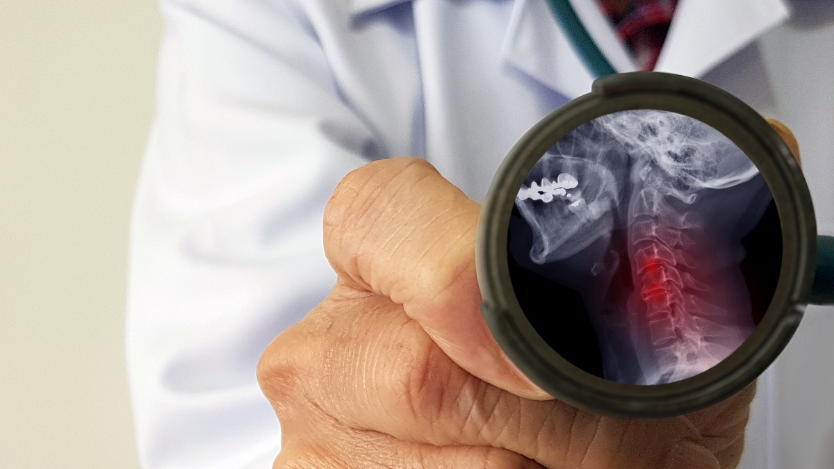
In fact, to diagnose cervical stenosis, the best study is the cervical Magnetic Resonance, which allows you to obtain all the information on the stage of the pathology, on the state of compression (anterior or posterior), on a possible condition of hypertrophy of the ligaments, on the presence or less than protrusions or herniated discs and to check for any spinal cord damage.
Cervical CT instead provides more information on the bone component.
X-rays of the spine allow you to analyze the vertebrae and to recognize any anatomical alterations affecting them (for example osteophytes).
MRI of the spine, on the other hand, analyzes in detail the soft and hard tissues of the anatomical area being investigated.
It is an effective test for verifying the consequences of aging and arthrosis at the vertebral level, and for diagnosing herniated discs.
Finally, the CT scan of the spine provides detailed three-dimensional images of the anatomical area of interest.
Myelography using X-rays and a radio-opaque contrast agent allows detailed examination of the spinal cord, spinal nerves, and other tissues in the spinal canal.
In practice, it allows you to accurately diagnose whether there is suffering from the spinal cord or spinal nerves due to cervical stenosis.
In addition, the physician may order neurophysiologic tests such as electromyography, somatosensory evoked potentials, and/or motor evoked potentials to document the degree of nerve distress in the nerve roots and spinal cord.
Treatment
In patients with a mild stenosis who therefore present mild symptoms, a conservative treatment is indicated, which includes physiotherapy sessions and the intake of drugs.
In particular, pain relievers and nonsteroidal anti-inflammatory drugs (NSAIDs) may be used temporarily for pain management (or corticosteroids when NSAIDs have not been successful) indicated by the doctor to relieve symptoms.
Functional rest, i.e. abstaining from all those activities or movements that evoke pain, is always advisable when dealing with a problem of this type, in particular when radiculopathy (i.e. friction of the lower nerve roots) is also found in the subject: in this case it is necessary that the neck is kept at rest and it may help to wear a soft orthopedic collar.
In the most serious cases, however, it is common to proceed with surgical decompression and cervical laminectomy, especially if the problem affects the spinal cord.
Surgery is in fact indicated in patients with severe stenoses, which also cause neurological disorders.
An anterior, posterior, or combined approach can be performed to decompress the spinal canal, depending on whether spinal cord compression prevails anteriorly, posteriorly, or both.
Through the anterior approach it is possible to remove the compression on the nerve root allowing to stabilize the cervical spine.

The posterior approach instead involves performing a posterior cervical midline incision of about 15 centimeters.
The paravertebral musculature is then bilaterally detached, reaching the laminae, then the surgeon evaluates whether to completely remove the laminae or proceed with an “open-door” laminotomy, thus cutting the laminae only on one side, thus allowing them to be opened en bloc.
In the combined approach, however, decompression is performed anteriorly and posteriorly.
Based on factors such as the age, the general conditions of the patient and the number of levels to be decompressed, it is also possible to evaluate performing the double operation in a single operating session, otherwise one proceeds in two successive stages.
Cervical stenosis, the prognosis
Generally, if the subject suffering from mild or moderate symptoms of cervical stenosis follows a conservative therapy, he does not suffer worsening.
The same thing happens for patients who need surgery; this means that surgical remedies are effective, albeit with a percentage of surgical complications.
Surgery is performed to prevent worsening or complications that lead to even more disabling neurological pictures, but, as regards the recovery of preoperative neurological disorders, there is always a percentage of uncertainty about patient expectations.
In cases where the stenosis has been complicated by damage to the marrow, unfortunately there is very little room for improvement but obviously this varies from patient to patient, from the characteristics of each one, from the quality and degree of adherence of the patient to the rehabilitation process.
Lumbar stenosis must be treated carefully because if it reaches very serious levels it can have repercussions on bowel and bladder function, leading to a loss of control of the anal and/or bladder sphincter.
Furthermore, always when it is very serious, it can be associated with paraplegia, i.e. a motor and/or sensory impairment of the lower part of the body, all complications that are difficult to regress after cervical decompression surgery.
Prevention of cervical stenosis
Preventing a condition like cervical stenosis is difficult, if not impossible when it is related to aging.
However, there are virtuous behaviors which, if scrupulously adopted, protect the spine and delay any pathologies affecting it, including the various forms of spinal stenosis. What to do?
- Adopt correct sitting posture.
- Lift heavy loads with the right technique (this also prevents herniated discs).
- Adopt a healthy lifestyle, i.e. a balanced diet (obesity and diabetes are proven risk factors for cervical stenosis).
- Not smoking.
Read Also
Emergency Live Even More…Live: Download The New Free App Of Your Newspaper For IOS And Android
What Is Cervicalgia? The Importance Of Correct Posture At Work Or While Sleeping
Cervicalgia: Why Do We Have Neck Pain?
Cervical Stenosis: Symptoms, Causes, Diagnosis And Treatment
Cervical Collar In Trauma Patients In Emergency Medicine: When To Use It, Why It Is Important
Headaches And Dizziness: It Could Be Vestibular Migraine
Migraine And Tension-Type Headache: How To Distinguish Between Them?
First Aid: Distinguishing The Causes Of Dizziness, Knowing The Associated Pathologies
Paroxysmal Positional Vertigo (BPPV), What Is It?
Cervical Dizziness: How To Calm It Down With 7 Exercises
Back Pain: Is It Really A Medical Emergency?
Posture, The Mistakes That Lead To Cervicalgia And Other Spinal Pains
Lumbago: What It Is And How To Treat It
Lumbar Puncture: What Is A LP?
General Or Local A.? Discover The Different Types
Intubation Under A.: How Does It Work?
How Does Loco-Regional Anaesthesia Work?
Are Anaesthesiologists Fundamental For Air Ambulance Medicine?
Epidural For Pain Relief After Surgery
Lumbar Puncture: What Is A Spinal Tap?
Lumbar Puncture (Spinal Tap): What It Consists Of, What It Is Used For
What Is Lumbar Stenosis And How To Treat It
Lumbar Spinal Stenosis: Definition, Causes, Symptoms, Diagnosis And Treatment