
Electrocardiogram: initial procedures, ECG electrode placement and some tips
If it is the patient’s first time performing an electrocardiogram (ECG), the rescuer, doctor, or nurse should explain to the patient – in words appropriate to his/her level of understanding – the steps and usefulness of the electrocardiogram
Before proceeding with the electrocardiogram, it is necessary to check
- whether there is any heart disease;
- which medications the patient is taking;
- any allergies to the materials used for the test (e.g. the gel used to facilitate the conduction of electrical signals);
- the presence of electronic devices that could alter the tracing (such as a pacemaker) or metal objects (such as necklaces and bracelets);
- the possible need for a trichotomy (if the patient has too much hair preventing the electrodes from adhering);
- the patient’s vital parameters, in particular blood pressure and heart rate.
The patient is asked
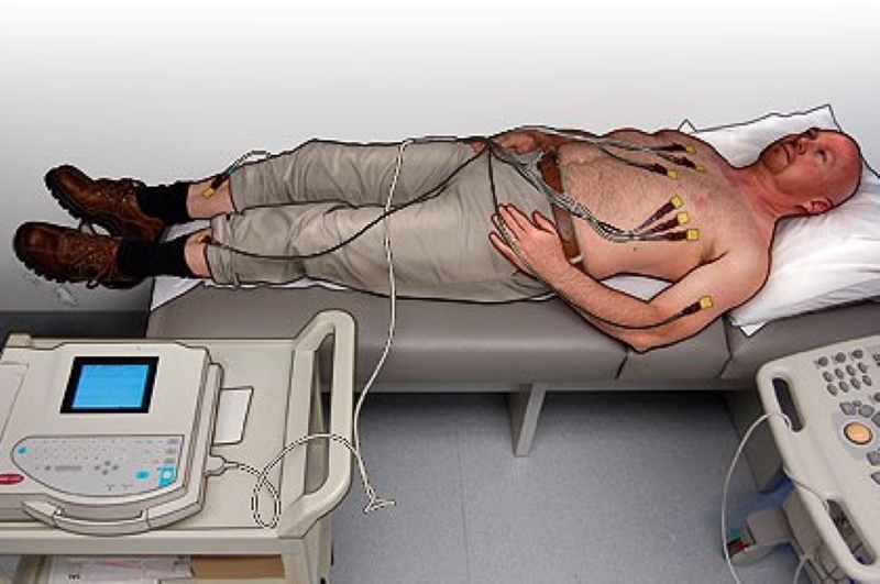
- to uncover their chest, ankles and wrists and to assume the supine position (belly up) on the couch, if possible;
- to relax and not to speak for a few minutes.
In the half-hour preceding the test, the patient should have avoided smoking and drinking coffee or alcohol.
Positioning the six electrodes
To position the six precordial leads:
- Identify where to place the V1 electrode (red): place it to the left of the patient; using the suprasternal fossa (dimple where the clavicles join, can be seen at the top of the sternum) as a reference, place the thumb of the left hand over it. Look for the first intercostal space with the index finger (stretching to the right and a little lower than the thumb), from here it will be automatic to find the second space with the middle finger, the third with the ring finger and finally with the little finger you will find the 4th right parasternal intercostal space: there V1 will be positioned.
- V2 (yellow): just to the left of V1, opposite side of the sternum you will find the 4th left parasternal intercostal space, there you should position V2. Important: the sternum is on average no more than 4 centimetres wide, so V1 and V2 should not be further apart than this.
- Position V3 (green) and V4 (brown): first position V4, locate the left clavicle and fix a point halfway along the bone. Then with the costal reference of V2 appreciate the 5th intercostal space with the finger technique. Now locate the point where you place V4 at the intersection of a line running down from the middle of the clavicle and finally meeting the 5th intercostal space. There you place the V4 electrode. V3 should be placed at the point midway between the line between V2 and V4 wherever this point is located, even if there are breasts present in women (does not prevent detection of the electrocardiac signal).
- Position V5 (black): locate the anterior axillary line, more or less where the axillary notch begins, and along this line, find the 5th intercostal space: there position V5.
- Position V6 (violet): locate the middle axillary line (about where the axillary hollow begins) and along this line, find the 5th intercostal space: there position V6. Take into account that V5 and V6 rise a little in relation to V4, because the ribs are concave upwards.
Electrocardiogram, positioning the four peripheral leads
To position the four peripheral leads, take the colour code into account:
- Red: right arm
- Black: right leg
- Green: left leg
- Yellow: left arm
Final checks
These are carried out before disconnecting the electrodes or, if there is a monitor, before printing:
- presence (and accuracy) of date and name;
- verification of the calibration signal and printing speed;
- quality of the trace:
- presence of all leads;
- stability of isoelectric line;
- absence of artefacts from tremors and alternating current;
- search for indications of incorrect electrode placement.
If the ECG electrodes are placed correctly, the trace of a healthy patient should have a result similar to this:
- Check for non-physiological electrocardiographic changes and if not, change the position of the electrodes, or bring the trace to the physician’s attention for evaluation and diagnosis.
Complications
Skin manifestations (e.g. reddening or itching of the skin) may sometimes occur after prolonged use of the electrodes due to the application time or skin sensitivity.
Advice
Keep the following tips and information in mind
- water is the best conductor of the electrical impulse, so to get a good ECG you should not use alcoholic disinfectants. It will look worse. If you have stockings or pantyhose, wet the gauze rather thoroughly (they should drip) and this will help conduct the electricity;
- one factor that improves recording is the placement of electrodes on bone planes, which conduct better than fat. For this reason, always choose the tibia and the back of the wrist for peripherals;
- positioning the electrodes correctly, because the detection of life-saving electrocardiographic abnormalities depends on this. Poor electrode placement can greatly affect the results of an ECG.
Electrocardiogram in a patient with an amputated limb
In patients who have had one or more limbs amputated, the electrode can be applied at any point on the limb stump or at the root of the limb.
Each limb is in fact considered, from an electrical point of view, to be a segment with low resistance, so that the electrical potential is essentially the same at all its points.
Of course, such a complex topic cannot be exhausted in a single article: more details will follow.

Read Also
Emergency Live Even More…Live: Download The New Free App Of Your Newspaper For IOS And Android
Performing The Cardiovascular Objective Examination: The Guide
What Is The Electrocardiogram (ECG)?
ECG: Waveform Analysis In The Electrocardiogram
What Is An ECG And When To Do An Electrocardiogram
ST-Elevation Myocardial Infarction: What Is A STEMI?
ECG First Principles From Handwritten Tutorial Video
ECG Criteria, 3 Simple Rules From Ken Grauer – ECG Recognize VT
The Patient’s ECG: How To Read An Electrocardiogram In A Simple Way

ECG: What P, T, U Waves, The QRS Complex And The ST Segment Indicate
Electrocardiogram (ECG): What It Is For, When It Is Needed
Stress Electrocardiogram (ECG): An Overview Of The Test
What Is The Dynamic Electrocardiogram ECG According To Holter?
Full Dynamic Electrocardiogram According To Holter: What Is It?
Cardiac Rhythm Restoration Procedures: Electrical Cardioversion
Cardiac Holter, The Characteristics Of The 24-Hour Electrocardiogram
Peripheral Arteriopathy: Symptoms And Diagnosis
Endocavitary Electrophysiological Study: What Does This Examination Consist Of?
Cardiac Catheterisation, What Is This Examination?
Echo Doppler: What It Is And What It Is For
Transesophageal Echocardiogram: What Does It Consist Of?
Paediatric Echocardiogram: Definition And Use
Heart Diseases And Alarm Bells: Angina Pectoris
Fakes That Are Close To Our Hearts: Heart Disease And False Myths
Sleep Apnoea And Cardiovascular Disease: Correlation Between Sleep And Heart
Myocardiopathy: What Is It And How To Treat It?
Venous Thrombosis: From Symptoms To New Drugs
Cyanogenic Congenital Heart Disease: Transposition Of The Great Arteries
Heart Rate: What Is Bradycardia?
Consequences Of Chest Trauma: Focus On Cardiac Contusion
Heart Murmur: What Is It And What Are The Symptoms?
Branch Block: The Causes And Consequences To Take Into Account
Cardiopulmonary Resuscitation Manoeuvres: Management Of The LUCAS Chest Compressor
Supraventricular Tachycardia: Definition, Diagnosis, Treatment, And Prognosis
Identifying Tachycardias: What It Is, What It Causes And How To Intervene On A Tachycardia
Myocardial Infarction: Causes, Symptoms, Diagnosis And Treatment
Aortic Insufficiency: Causes, Symptoms, Diagnosis And Treatment Of Aortic Regurgitation
Congenital Heart Disease: What Is Aortic Bicuspidia?
Atrial Fibrillation: Definition, Causes, Symptoms, Diagnosis And Treatment
Ventricular Fibrillation Is One Of The Most Serious Cardiac Arrhythmias: Let’s Find Out About It
Atrial Flutter: Definition, Causes, Symptoms, Diagnosis And Treatment
What Is Echocolordoppler Of The Supra-Aortic Trunks (Carotids)?
What Is The Loop Recorder? Discovering Home Telemetry