
Stress Electrocardiogram (ECG): an overview of the test
What is a stress electrocardiogram (ECG)? The electrocardiogram or ECG is a diagnostic test performed to record the electrical activity of the heart, so that its general state of health can be assessed and various cardiac conditions, sinus rhythm arrhythmias or heart disease can be detected
The investigation is carried out using an instrument called an electrocardiograph, which is able to record the heart function and reproduce it graphically in the form of a trace.
Depending on the case, a cardiologist may prescribe different types of ECG, which differ mainly according to the specific function and how they are performed: the standard or resting ECG, the dynamic ECG according to Holter, and the exercise ECG or ergometric test.
Resting electrocardiogram: used to record cardiac function under normal conditions
It involves placing 12 to 15 electrodes on the patient’s chest, arms and legs; the test usually lasts a few minutes, during which all that is required is to lie still, breathe regularly and avoid talking or moving.
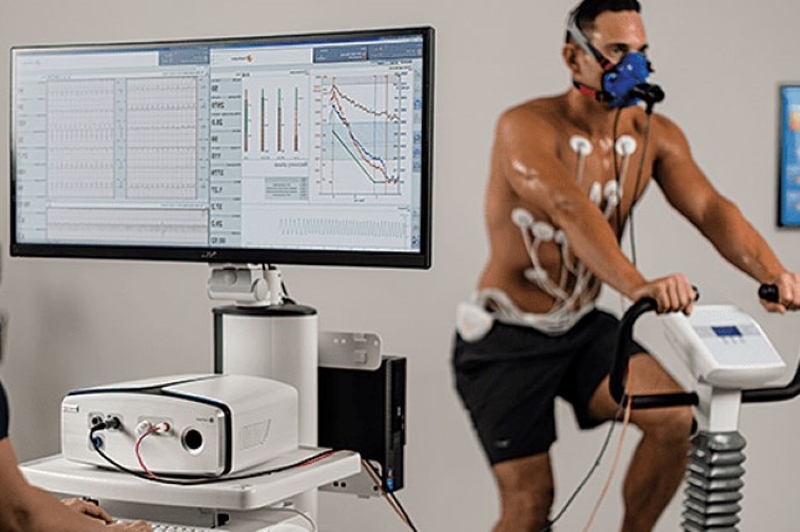
Stress electrocardiogrph: assesses ECG changes during physical activity
It is also used to assess cardiac behaviour when subjected to exertion; after placing the electrodes, the patient is generally asked to pedal on an exercise bike or run on a treadmill for 5 to 20 minutes to take the measurement.
Dynamic electrocardiogram according to Holter:
This is performed to monitor heart function over a longer period of time, usually 24 – 48 hours; it is usually prescribed to diagnose arrhythmias of a discontinuous character, which would not be detected by a standard ECG.
To perform the examination, electrodes are attached to a portable device that records cardiac activity and allows the tracing to be extrapolated later.
Why is the stress electrocardiogram performed?
The stress test is used to monitor cardiac function when subjected to intense physical activity: it allows variations in the number of beats and alterations in cardiac rhythm to be detected; the examination involves performing simple physical exercises, or the administration of specific drugs suitable for simulating the effects of stress.
At the physician’s request, oxygen consumption may be prescribed during the exercise ECG in order to assess the change in blood pressure levels and monitor the pulmonary response related to respiratory function.
The exercise ECG makes it possible to examine the reactions of the cardiovascular system and diagnose any abnormalities that would not be detected under normal conditions
In particular, it is indicated for the in-depth study of
- ischaemic heart disease
- arrhythmias;
- arterial hypertension;
- angina pectoris;
- assessing the effectiveness of interventions such as angioplasty and bypass;
- in some cases, this investigation is prescribed to assess the effects of certain drugs on the heart.
Stress Electrocardiogram (ECG): How should I prepare?
The ECG is a non-invasive examination that poses no risk to the patient and requires no special preparation.
Some useful precautions may be
- wear comfortable clothing that allows freedom of movement;
- perform the test on an empty stomach for at least 2-3 hours;
- avoid alcohol or caffeine;
- abstaining from smoking;
- do not engage in strenuous sport in the 4-5 hours preceding the test;
- take any medication regularly, unless otherwise instructed by your doctor.
*This is only indicative information: it is therefore necessary to contact the facility where the examination is being performed to obtain specific information on the preparation procedure.
Read Also:
Emergency Live Even More…Live: Download The New Free App Of Your Newspaper For IOS And Android
ECG: Waveform Analysis In The Electrocardiogram
What Is An ECG And When To Do An Electrocardiogram
ST-Elevation Myocardial Infarction: What Is A STEMI?
ECG First Principles From Handwritten Tutorial Video
ECG Criteria, 3 Simple Rules From Ken Grauer – ECG Recognize VT
Defibrillator: What It Is, How It Works, Price, Voltage, Manual And External
The Patient’s ECG: How To Read An Electrocardiogram In A Simple Way
ECG: What P, T, U Waves, The QRS Complex And The ST Segment Indicate
Endocavitary Electrophysiological Study: What Does This Examination Consist Of?
Head Up Tilt Test, How The Test That Investigates The Causes Of Vagal Syncope Works

What Is Ischaemic Heart Disease And Possible Treatments
Percutaneous Transluminal Coronary Angioplasty (PTCA): What Is It?
Ischaemic Heart Disease: What Is It?
EMS: Pediatric SVT (Supraventricular Tachycardia) Vs Sinus Tachycardia
Paediatric Toxicological Emergencies: Medical Intervention In Cases Of Paediatric Poisoning
Valvulopathies: Examining Heart Valve Problems
What Is The Difference Between Pacemaker And Subcutaneous Defibrillator?
Heart Disease: What Is Cardiomyopathy?
Inflammations Of The Heart: Myocarditis, Infective Endocarditis And Pericarditis
Heart Murmurs: What It Is And When To Be Concerned
Clinical Review: Acute Respiratory Distress Syndrome
Botallo’s Ductus Arteriosus: Interventional Therapy
Heart Valve Diseases: An Overview
Cardiomyopathies: Types, Diagnosis And Treatment
First Aid And Emergency Interventions: Syncope
Tilt Test: What Does This Test Consist Of?
Cardiac Syncope: What It Is, How It Is Diagnosed And Who It Affects
New Epilepsy Warning Device Could Save Thousands Of Lives
Understanding Seizures And Epilepsy
First Aid And Epilepsy: How To Recognise A Seizure And Help A Patient
Neurology, Difference Between Epilepsy And Syncope
Positive And Negative Lasègue Sign In Semeiotics
Wasserman’s Sign (Inverse Lasègue) Positive In Semeiotics
Positive And Negative Kernig’s Sign: Semeiotics In Meningitis
Lithotomy Position: What It Is, When It Is Used And What Advantages It Brings To Patient Care
Trendelenburg (Anti-Shock) Position: What It Is And When It Is Recommended
Prone, Supine, Lateral Decubitus: Meaning, Position And Injuries
Stretchers In The UK: Which Are The Most Used?
Does The Recovery Position In First Aid Actually Work?
Reverse Trendelenburg Position: What It Is And When It Is Recommended
Drug Therapy For Typical Arrhythmias In Emergency Patients
Canadian Syncope Risk Score – In Case Of Syncope, Patients Are Really In Danger Or Not?