
Capnography in ventilatory practice: why do we need a capnograph?
Ventilation must be performed correctly, sufficient monitoring is necessary: the capnographer plays a precise role in this
The capnograph in the mechanical ventilation of the patient
If necessary, mechanical ventilation in the prehospital phase must be performed correctly and with comprehensive monitoring.
It is important not only to get the patient to hospital, but also to ensure a high chance of recovery, or at least not to aggravate the severity of the patient’s condition during transport and care.
The days of simpler ventilators with minimal settings (frequency-volume) are a thing of the past.
Most patients requiring mechanical ventilation have partially preserved spontaneous breathing (bradypnoea and hypoventilation), which lies in the middle of the ‘range’ between complete apnoea and spontaneous breathing, where oxygen inhalation is sufficient.
ALV (Adaptive lung ventilation) in general should be normoventilation: hypoventilation and hyperventilation are both harmful.
The effect of inadequate ventilation on patients with acute brain pathology (stroke, head trauma, etc.) is particularly harmful.
Hidden enemy: hypocapnia and hypercapnia
It is well known that breathing (or mechanical ventilation) is necessary to supply the body with oxygen O2 and remove carbon dioxide CO2.
The damage of a lack of oxygen is obvious: hypoxia and brain damage.
Excess O2 can damage the epithelium of the airways and the alveoli of the lungs, however, when using an oxygen concentration (FiO2) of 50% or less, there will be no significant damage from ‘hyperoxygenation’: the unassimilated oxygen will simply be removed with exhalation.
CO2 excretion does not depend on the composition of the supplied mixture and is determined by the minute ventilation value MV (frequency, f x tidal volume, Vt); the thicker or deeper the breath, the more CO2 is excreted.
With a lack of ventilation (‘hypoventilation’) – bradypnoea/superficial breathing in the patient himself or mechanical ventilation ‘lacks’ hypercapnia (excess CO2) progresses in the body, in which there is a pathological expansion of cerebral vessels, an increase in intracranial pressure, cerebral oedema and its secondary damage.
But with excessive ventilation (tachypnoea in a patient or excessive ventilation parameters), hypocapnia is observed in the body, in which there is pathological narrowing of the cerebral vessels with ischaemia of its sections, and thus also secondary brain damage, and respiratory alkalosis also aggravates the severity of the patient’s condition. Therefore, mechanical ventilation should not only be ‘anti-hypoxic’, but also ‘normocapnic’.
There are methods for theoretically calculating mechanical ventilation parameters, such as Darbinyan’s formula (or other corresponding ones), but they are indicative and may not take into account the patient’s actual condition, for example.
Why a pulse oximeter is not sufficient
Of course, pulse oximetry is important and forms the basis of ventilation monitoring, but SpO2 monitoring is not sufficient, there are a number of hidden problems, limitations or dangers, namely: In the situations described, the use of a pulse oximeter often becomes impossible.
– When using oxygen concentrations above 30% (usually FiO2 = 50% or 100% is used with ventilation), reduced ventilation parameters (rate and volume) may be sufficient to maintain “normoxia” as the amount of O2 delivered per respiratory act increases. Therefore, the pulse oximeter will not show hidden hypoventilation with hypercapnia.
– The pulse oximeter does not show harmful hyperventilation in any way, constant SpO2 values of 99-100% falsely reassure the physician.
– The pulse oximeter and the saturation indicators are very inert, due to the supply of O2 in the circulating blood and the physiological dead space of the lungs, as well as due to the averaging of readings over a time interval on the pulse oximeter-protected transport pulse, in the event of an emergency event (circuit disconnection, lack of ventilation parameters, etc.) n.) saturation does not decrease immediately, whereas a quicker response from the physician is required.
– The pulse oximeter gives incorrect SpO2 readings in case of carbon monoxide (CO) poisoning due to the fact that the light absorption of oxyhaemoglobin HbO2 and carboxyhaemoglobin HbCO is similar, monitoring in this case is limited.
Use of the capnograph: capnometry and capnography
Additional monitoring options that save the patient’s life.
A valuable and important addition to the control of the adequacy of mechanical ventilation is the constant measurement of the CO2 concentration (EtCO2) in the exhaled air (capnometry) and a graphical representation of the cyclicity of CO2 excretion (capnography).
The advantages of capnometry are:
– Clear indicators in any haemodynamic state, even during CPR (at critically low blood pressure, monitoring is done via two channels: ECG and EtCO2)
– Instant change of indicators for any events and deviations, e.g. when the respiratory circuit is disconnected
– Assessment of initial respiratory status in an intubated patient
– Real-time visualisation of hypo- and hyperventilation
Further features of capnography are extensive: airway obstruction is shown, the patient’s attempts to breathe spontaneously with the need to deepen anaesthesia, cardiac oscillations on the chart with tachyarrhythmia, a possible rise in body temperature with an increase in EtCO2 and much more.
Main objectives of using a capnograph in the prehospital phase
Monitoring the success of tracheal intubation, especially in situations of noise and difficulty of auscultation: the normal programme of cyclic CO2 excretion with good amplitude will never work if the tube is inserted into the oesophagus (however, auscultation is necessary to control ventilation of the two lungs)
Monitoring the restoration of spontaneous circulation during CPR: metabolism and CO2 production increase significantly in the ‘resuscitated’ organism, a ‘jump’ appears on the capnogram and the visualisation does not worsen with cardiac compressions (unlike the ECG signal)
General control of mechanical ventilation, especially in patients with brain damage (stroke, head injury, convulsions, etc.)
Measurement “in the main flow” (MAINSTREAM) and “in the lateral flow” (SIDESTREAM).
Capnographs are of two technical types, when measuring EtCO2 ‘in the main stream’ a short adapter with side holes is placed between the endotracheal tube and the circuit, a U-shaped sensor is placed on it, the passing gas is scanned and the determined EtCO2 is measured.
When measuring ‘in a lateral flow’, a small portion of gas is taken from the circuit through a special hole in the circuit by the suction compressor, is fed through a thin tube into the body of the capnograph, where the EtCO2 is measured.
Several factors influence the accuracy of the measurement, such as the concentration of O2 and moisture in the mixture and the measuring temperature. The sensor must be preheated and calibrated.
In this sense, the sidestream measurement appears to be more accurate, as it reduces the influence of these distorting factors in practice, however.
Portability, 4 versions of the capnograph:
- as part of a bedside monitor
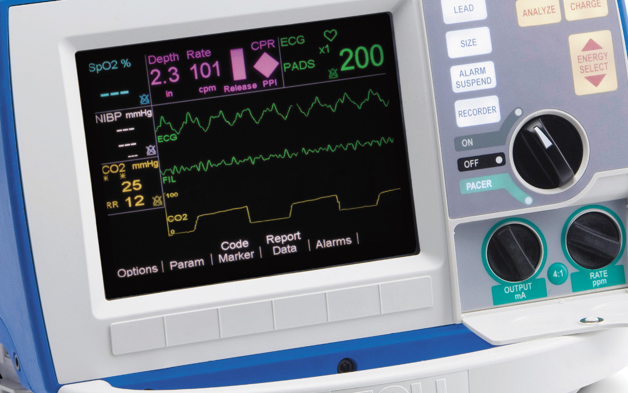
- as part of a multifunctional defibrillator
- a mini-nozzle on the circuit (‘device is in the sensor, no wire’)
- a portable pocket device (‘body + sensor on the wire’).
Usually, when referring to capnography, the EtCO2 monitoring channel is understood as part of a multifunctional ‘bedside’ monitor; in the ICU, it is permanently fixed on the equipment shelf.
Although the monitor stand is removable and the capnograph monitor is powered by a built-in battery, it is still difficult to use it when moving to the flat or between the rescue vehicle and the intensive care unit, due to the weight and size of the monitor case and the impossibility of attaching it to a patient or to a waterproof stretcher, on which the transport from the flat was mainly carried out.
A much more portable instrument is needed.
Similar difficulties are encountered when using a capnograph as part of a professional multifunctional defibrillator: unfortunately, almost all of them still have a large size and weight, and in reality do not allow, for example, such a device to be comfortably placed on a waterproof stretcher next to the patient when descending stairs from a high floor; even during operation, confusion often occurs with a large number of wires in the device.
Read Also

Emergency Live Even More…Live: Download The New Free App Of Your Newspaper For IOS And Android
What Is Hypercapnia And How Does It Affect Patient Intervention?
Ventilatory Failure (Hypercapnia): Causes, Symptoms, Diagnosis, Treatment
How To Choose And Use A Pulse Oximeter?
Equipment: What Is A Saturation Oximeter (Pulse Oximeter) And What Is It For?
Basic Understanding Of The Pulse Oximeter
Three Everyday Practices To Keep Your Ventilator Patients Safe
Medical Equipment: How To Read A Vital Signs Monitor
Ambulance: What Is An Emergency Aspirator And When Should It Be Used?
Ventilators, All You Need To Know: Difference Between Turbine Based And Compressor Based Ventilators
Life-Saving Techniques And Procedures: PALS VS ACLS, What Are The Significant Differences?
The Purpose Of Suctioning Patients During Sedation
Supplemental Oxygen: Cylinders And Ventilation Supports In The USA
Basic Airway Assessment: An Overview
Ventilator Management: Ventilating The Patient
Emergency Equipment: The Emergency Carry Sheet / VIDEO TUTORIAL
Defibrillator Maintenance: AED And Functional Verification
Respiratory Distress: What Are The Signs Of Respiratory Distress In Newborns?
EDU: Directional Tip Suction Catheter
Suction Unit For Emergency Care, The Solution In A Nutshell: Spencer JET
Airway Management After A Road Accident: An Overview
Tracheal Intubation: When, How And Why To Create An Artificial Airway For The Patient
What Is Transient Tachypnoea Of The Newborn, Or Neonatal Wet Lung Syndrome?
Traumatic Pneumothorax: Symptoms, Diagnosis And Treatment
Diagnosis Of Tension Pneumothorax In The Field: Suction Or Blowing?
Pneumothorax And Pneumomediastinum: Rescuing The Patient With Pulmonary Barotrauma
ABC, ABCD And ABCDE Rule In Emergency Medicine: What The Rescuer Must Do
Multiple Rib Fracture, Flail Chest (Rib Volet) And Pneumothorax: An Overview
Internal Haemorrhage: Definition, Causes, Symptoms, Diagnosis, Severity, Treatment
Assessment Of Ventilation, Respiration, And Oxygenation (Breathing)
Oxygen-Ozone Therapy: For Which Pathologies Is It Indicated?
Difference Between Mechanical Ventilation And Oxygen Therapy
Hyperbaric Oxygen In The Wound Healing Process
Venous Thrombosis: From Symptoms To New Drugs
What Is Intravenous Cannulation (IV)? The 15 Steps Of The Procedure
Nasal Cannula For Oxygen Therapy: What It Is, How It Is Made, When To Use It
Nasal Probe For Oxygen Therapy: What It Is, How It Is Made, When To Use It
Oxygen Reducer: Principle Of Operation, Application
How To Choose Medical Suction Device?
Holter Monitor: How Does It Work And When Is It Needed?
What Is Patient Pressure Management? An Overview
Head Up Tilt Test, How The Test That Investigates The Causes Of Vagal Syncope Works
Cardiac Syncope: What It Is, How It Is Diagnosed And Who It Affects
Cardiac Holter, The Characteristics Of The 24-Hour Electrocardiogram