
Diagnostic Imaging in Oncology
In oncology, CT and MRI are the two most widely used digital imaging methods in the approach to the neoplastic patient
These two diagnostic tools find indication in the different phases of the natural history of patients from tumour recognition to the clinical manifestation, to the differential diagnosis with other forms of non-developmental pathology, to the evaluation of the response to treatments as well as, obviously, to the definition when obtainable of the cure.
Computed axial tomography (CT) in oncology
It makes use of ionising radiation to construct diagnostic images and provides an anatomical representation of the human body based on the different degree to which tissues filter the radiation beam.
A representation of normal and pathological human body anatomy is thus obtained on axial cut sections.
The most recent generation of CT equipment (spiral CT) makes it possible to acquire images of great detail, with the possibility of obtaining reconstructions in the various possible anatomical planes, in addition to the axial one, without losing image quality.
In the field of oncology, the spread of such equipment has opened up new scenarios for quantifying the vascularisation of diseased tissue, addressing the problems inherent in assessing the activity of new antineoplastic drugs based on the destruction of the neoplastic tissue’s supply vessels (anti-neoangiogenetic drugs).
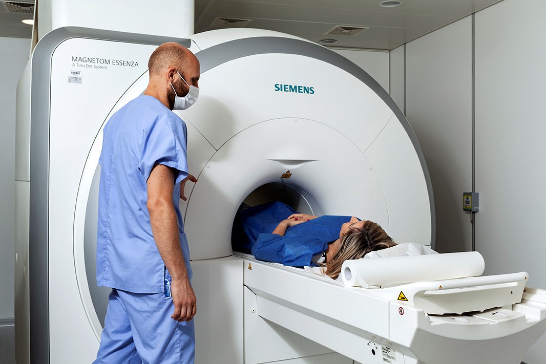
Magnetic Resonance Imaging (MRI) in Oncology
Directly produces images in different scan planes.
This diagnostic tool, which originated in the hands of chemists for structural studies of matter and only came to medicine at the end of the last century, uses the energy of very high magnetic fields in conjunction with radiofrequency sources to construct the images.
Starting from a biochemical structural basis enriches MRI images and makes them suitable for not only morphological but also structural tissue definition. In other words, with MRI, the tumour is no longer seen only as an ‘extraneous’ space-occupying mass in an anatomical territory, but also as an area of altered signal in the context of an organ or tissue without changes in its volume or profile.
The quality of MRI images is directly proportional to the magnetic field strength, which generally varies in radiological diagnostic applications from 0.5 to 1.5 Tesla (unit of measurement of field strength).
The 3 Tesla MRI is capable of obtaining images within a few seconds (so much so that they can be acquired during respiratory apnoea), with the advantage of reducing the patient’s overall time in the machine.
Finally, the new magnet is particularly compact and the width of the ‘tube’ in which the patient is positioned is particularly wide, so as to minimise the negative impact on patients suffering from the forced position and claustrophobia.
Read Also
Emergency Live Even More…Live: Download The New Free App Of Your Newspaper For IOS And Android
Fusion Prostate Biopsy: How The Examination Is Performed
Spinal Biopsy: What It Is, How It Is Performed And What Risks It Presents

Echo- And CT-Guided Biopsy: What It Is And When It Is Needed
What Is Needle Aspiration (Or Needle Biopsy Or Biopsy)?
What Is Echocolordoppler Of The Supra-Aortic Trunks (Carotids)?
What Is The Loop Recorder? Discovering Home Telemetry
Cardiac Holter, The Characteristics Of The 24-Hour Electrocardiogram
Peripheral Arteriopathy: Symptoms And Diagnosis
Endocavitary Electrophysiological Study: What Does This Examination Consist Of?
Cardiac Catheterisation, What Is This Examination?
Echo Doppler: What It Is And What It Is For
Transesophageal Echocardiogram: What Does It Consist Of?
Venous Thrombosis: From Symptoms To New Drugs
Echotomography Of Carotid Axes
What Is A Liver Biopsy And When Is It Performed?
Abdominal Ultrasound: How It Is Performed And What It Is Used For
What Is Retinal Fluorangiography And What Are The Risks?
Echodoppler: What It Is And When To Perform It
Biopsy: What It Is And When It Is Performed