
What is retinal fluorangiography and what are the risks?
Retinal fluorangiography (FAG), or fluorescein angiography, is used to diagnose vascular diseases of the eye and in particular those affecting the retina, including diabetic retinopathy, age-related macular degeneration, and vascular occlusions
A painless and quick test, it is carried out by means of a simple photograph, without contact with the eye.
It is an increasingly frequent investigation, also in view of the rising average age and the increasing prevalence of diseases such as diabetes.
What is the retina
The retina is a transparent membrane located inside the eyeball and is responsible for transforming light signals from outside into images.
In detail, it is made up of 2 zones
- the macula, i.e. the central area that allows objects, movements and faces to be distinguished;
- the optic papilla, i.e. the area where the optic nerve (nerve that transmits visual information from the retina to the brain) meets the retina.
What is fluorangiography
Retinal fluorangiography is a diagnostic imaging technique that makes it possible to visualise and photograph in detail, thanks to the use of a particular dye, the blood vessels of the ocular fundus and highlight any circulatory pathologies affecting the retina, the most frequent of which are:
- diabetic retinopathy
- age-related degeneration of the macula;
- vascular occlusions.
How the test is performed
The first phase of the test involves:
- administration of a dye contrast medium, called fluorescein;
- dilation of the pupils with a mydriatic eye drops.
The contrast medium, injected into a vein in the arm, immediately enters the venous circulation and diffuses through the body in a few seconds, reaching the blood vessels of the eye.
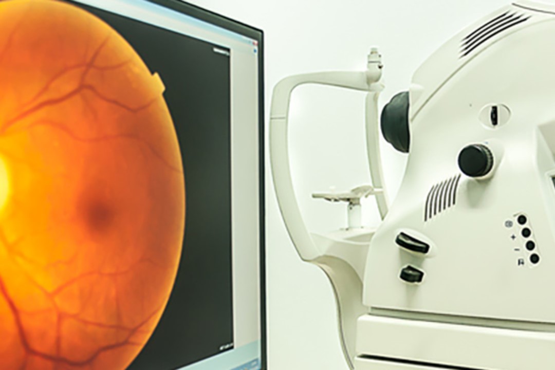
Immediately afterwards, the actual test is carried out with an instrument called a fluorangiograph that illuminates the fundus of the eye with a light of a suitable wavelength, such as blue light, which can stimulate the contrast medium to emit fluorescence.
The instrument’s optical system detects the fluorescence emitted by the blood vessels and uses a computer programme to reconstruct an accurate and detailed digitised image of the vessels analysed and the blood flow within them.
The test takes less than half an hour to perform and does not require the use of contrast medium.
What fluorangiography is used for
Based on the digitised images of the retinal blood vessels obtained with retinal fluorangiography, the ophthalmologist is able to
- diagnose any eye diseases of vascular origin;
- plan possible medical therapy or photocoagulative laser treatment;
- guide the execution of photocoagulative laser treatment;
- monitor the clinical evolution of the pathology.
Preliminary tests to be performed
Before undergoing fluorangiography, in order to perform it in complete safety, it is necessary to undergo certain tests, especially in patients with known pathologies (heart disease, renal insufficiency, etc.) or with declared allergies to drugs and/or food.
In detail, the tests required are:
- blood tests (creatinine for the assessment of renal function, which is necessary for the proper disposal of the dye used as a contrast agent, blood glucose, complete blood count, etc.);
- electrocardiogram if the patient has a pre-existing heart condition.
If the patient is allergic to drugs, it is useful to perform a so-called desensitising prophylaxis, which involves taking
- cortisone tablets (the evening before the test and in the morning);
- ketotifen tablets (the morning of the test).
Fluorescein angiography is a minimally invasive test and usually has no major side effects
The most common side effects include
- darkened or slightly coloured vision for a few minutes
- yellowish discolouration of the skin for a few hours;
- yellowish or dark orange urine for up to 24 hours after the test;
- burning sensation at the point on the arm where the injection was made, usually caused by some dye escaping from the vein.
Allergic reactions to fluorescein are very rare and may manifest as:
- allergic dermatitis
- itching;
- difficulty breathing.
These reactions are usually treated with an antihistamine or an oral or injectable cortisone, depending on the severity of the symptoms.
Allergic reactions are rare events, but should never be underestimated.
Precautions after the fluorangiography test
Hypersensitivity to light may occur after the test due to dilation of the pupils, which is why it is advisable to wear dark sunglasses.

Since pupil dilation can also lead to blurred vision, the patient should be accompanied.
Read Also
Emergency Live Even More…Live: Download The New Free App Of Your Newspaper For IOS And Android
Red Eyes: What Can Be The Causes Of Conjunctival Hyperemia?
Autoimmune Diseases: The Sand In The Eyes Of Sjögren’s Syndrome
How To Prevent Dry Eyes During Winter: Tips
Corneal Abrasions And Foreign Bodies In The Eye: What To Do? Diagnosis And Treatment
Covid, A ‘Mask’ For The Eyes Thanks To Ozone Gel: An Ophthalmic Gel Under Study
Dry Eyes In Winter: What Causes Dry Eye In This Season?
What Is Aberrometry? Discovering The Aberrations Of The Eye
Stye Or Chalazion? The Differences Between These Two Eye Diseases
Eye For Health: Cataract Surgery With Intraocular Lenses To Correct Visual Defects
Cataract: Symptoms, Causes And Intervention
Inflammations Of The Eye: Uveitis
Corneal Keratoconus, Corneal Cross-Linking UVA Treatment
Myopia: What It Is And How To Treat It
Presbyopia: What Are The Symptoms And How To Correct It
Nearsightedness: What It Myopia And How To Correct It
Blepharoptosis: Getting To Know Eyelid Drooping
Lazy Eye: How To Recognise And Treat Amblyopia?
What Is Presbyopia And When Does It Occur?
Presbyopia: An Age-Related Visual Disorder
Blepharoptosis: Getting To Know Eyelid Drooping
Rare Diseases: Von Hippel-Lindau Syndrome
Rare Diseases: Septo-Optic Dysplasia
Diseases Of The Cornea: Keratitis
Heart Attack, Prediction And Prevention Thanks To Retinal Vessels And Artificial Intelligence
Eye Care And Prevention: Why It Is Important To Have An Eye Examination
Dry Eye Syndrome: Symptoms, Causes And Remedies
Maculopathy: Symptoms And How To Treat It
Dry Eye Syndrome: How To Protect Your Eyes From PC Exposure
Reddening Of The Eyes: What Diseases Are Linked To Eye Redness?