
Electromyography: what is it and what is it used for when treating nerve disorders?
Let’s talk about electromyography: many people, having reached a certain age, experience motor disturbances. They are unable to grasp objects, can no longer walk properly, or have difficulty performing normal everyday gestures
Many people who perform a certain type of movement with a certain frequency (e.g. pianists), may experience even very painful disorders in the joints used to perform that movement.
Many people cannot understand what is happening to their body, but they experience pain, discomfort, tingling, and an inability to perform certain movements.
All these people, as well as those who have suffered a trauma or injury, need to undergo a special test to verify the functionality of the affected parts, this test is called electromyography, let’s see what it is.
What is electromyography
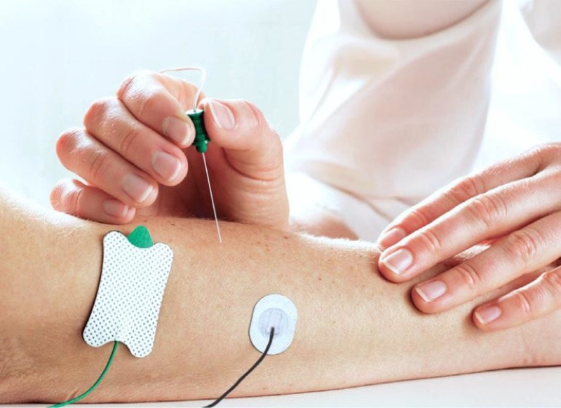
Electromyography is the diagnostic test used to assess the function of nerves and muscles, thus detecting whether there is neuropathy or myopathy.
It is therefore a functional test, i.e. it investigates the functionality of the muscles or nerves being tested.
This functionality can be detected by means of needle electromyography, i.e. using thin needles of different lengths depending on the muscle to be assessed, which are introduced into the muscle in order to record its activity.
Then there is the conduction velocity test, or electroneurography (ENG), which is used to assess the information conduction capacity of nerves, using electrical stimuli that are applied with electrodes placed on the skin, in some cases subcutaneously with small needles.
These tests are different from CT scans and ultrasound scans which are, instead, morphological tests.
Thus, in essence, electromyography serves to have an evaluation of the functionality of muscles and nerves, and obviously to highlight any neurological problems.
Often a strong tingling sensation, especially in the arms, is misinterpreted and one quickly goes to the doctor for fear that it is a heart attack, but then it turns out that it could be some kind of nerve or muscle disorder.
When should this test be done?
Electromyography is prescribed when there are conditions that may lead one to suspect neurological or peripheral damage, or following a trauma or injury in order to assess the patient’s condition.
Among the main reasons why the doctor requires such a test are intense and continuous tingling, numbness, muscle weakness, muscle paralysis, pain, muscle spasms.
It therefore serves to diagnose various pathologies, some of which can be resolved completely and are not disabling, and others that can still be resolved quite effectively.
These include carpal tunnel syndromes, ulnar groove syndromes, rasal tunnel syndromes, but also polyneuropathies, radiocolopathies, amniotrophic lateral sclerosis (ALS), muscular diseases such as myopathies or myositis, myasthenia gravis, plexopathies, Guyon’s syndrome and so on.
There are various nerve and muscle affections, diseases that can affect, even severely, movement and thus the normal performance of everyday actions, rendering the affected person incapable or partially incapacitated.
Even in the case of trauma and injuries, muscles and nerves can be damaged, but thanks to electromyography it is possible to understand the degree of damage and hypothesise the therapy for partial or total recovery.
Needle electromyography is a moderately invasive test and cannot be performed by everyone
Pacemaker wearers, for example, should only undergo the test after careful evaluation of pros and cons by their doctor.
There are no studies showing that the functioning of the pacemaker is altered during the conduction velocity test, but it should be assessed on a case-by-case basis.
Patients at risk of bleeding, e.g. those suffering from haemophilia, should undergo the test after careful evaluation.
Slight bleeding and bruising are normal after the test.
The condition of patients with lymphoedema and brain stimulators should also be assessed.
There are no contraindications for carrying out the test during pregnancy, but the doctor should be informed.
How to prepare for the test

Before the test it is best not to apply creams or oils to the body, at least a day or two before, while nail varnish or false nails are not a problem.
Bringing previous tests is always a good rule.
The test itself is not painful, however it depends on the pain sensitivity of each subject.
If the complete test is to be performed, one will have to get used to the idea of needles and small electric shocks.
Read Also
Emergency Live Even More…Live: Download The New Free App Of Your Newspaper For IOS And Android
Electromyography (EMG), What It Assesses And When It Is Done
Echo-Colour Doppler Of The Renal Arteries: What Does It Consist Of?
Echodoppler: What It Is And When To Perform It
Echo Doppler: What It Is And What It Is For
Fusion Prostate Biopsy: How The Examination Is Performed
What Is Needle Aspiration (Or Needle Biopsy Or Biopsy)?
What Is Echocolordoppler Of The Supra-Aortic Trunks (Carotids)?
What Is The Loop Recorder? Discovering Home Telemetry
Cardiac Holter, The Characteristics Of The 24-Hour Electrocardiogram
Peripheral Arteriopathy: Symptoms And Diagnosis
Endocavitary Electrophysiological Study: What Does This Examination Consist Of?
Cardiac Catheterisation, What Is This Examination?
Transesophageal Echocardiogram: What Does It Consist Of?
Venous Thrombosis: From Symptoms To New Drugs
Echotomography Of Carotid Axes
Echo- And CT-Guided Biopsy: What It Is And When It Is Needed
Echo-Doppler Of Vessels: Characteristics And Limitations Of The Method