
What is coronary angiography?
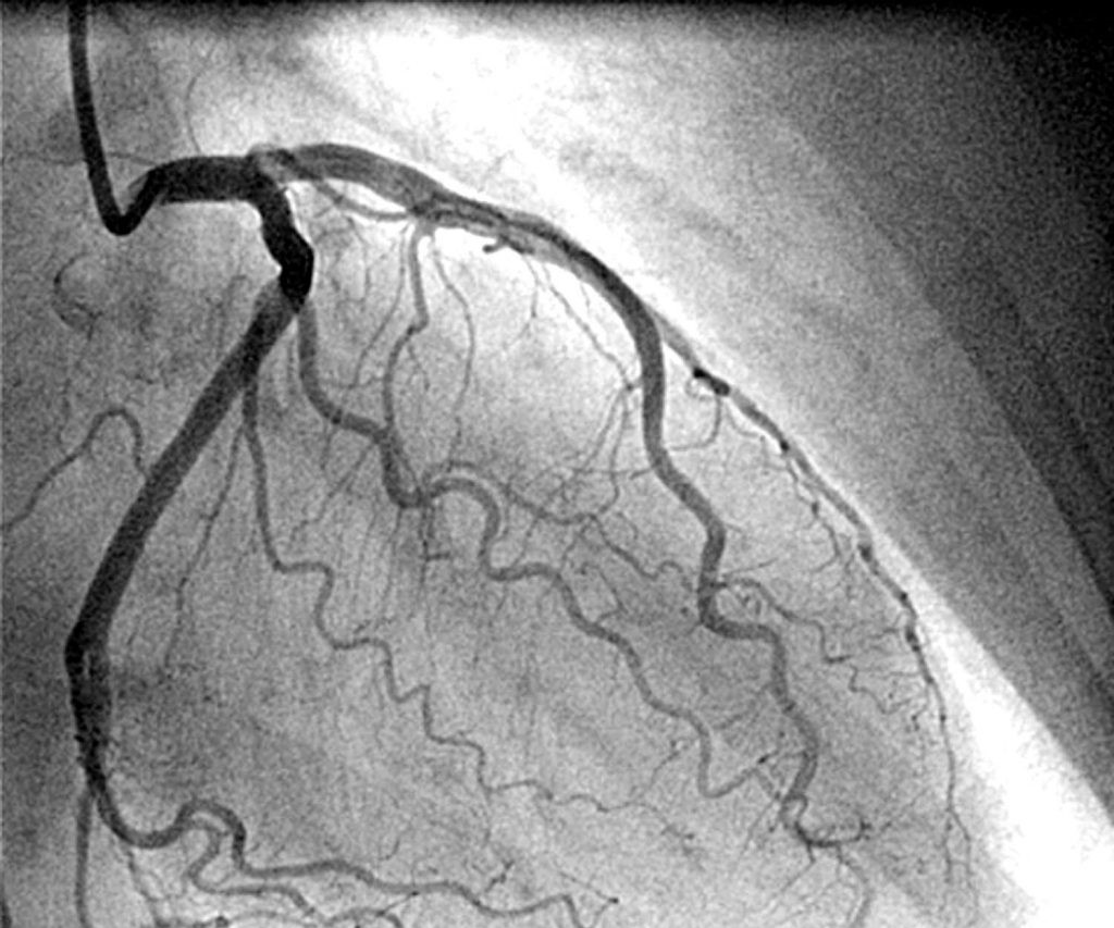
Coronary angiography is a radiological examination that allows you to view images of the coronary arteries, the arterial vessels that surround the heart and carry blood to the heart muscle
It is a diagnostic procedure that is carried out through the introduction of a contrast medium into the bloodstream, which is useful for making the coronary arteries visible to the machine.
Coronagraphy involves the introduction of a catheter, a thin, flexible tube that is advanced into the blood vessels to the point where it must release the contrast fluid.
Thanks to the advent of digital technologies, today it is possible to obtain images of the circulatory function by minimizing the use of the contrast medium.
What is coronary angiography for?
Coronagraphy is a test indicated to evaluate heart function.
It allows you to establish whether the coronary arteries are free (patent) or obstructed by clots, narrowing (stenosis) or cholesterol plaques (atheromas).
It is indicated when the patient reports:
- Chest pain (angina pectoris) or anginal pain in the arm
- A genetic defect from birth (congenital heart disease)
- Heart valve defects
- Heart failure
- Trauma
Angiography allows you to schedule surgery
For example, it can precede or be associated with angioplasty, which involves the introduction of a stent to restore flow in an occluded vessel.
It is useful for assessing possible complications of surgery.
It is also a diagnostic method to monitor the results of an intervention (follow up) such as in the case of a bypass.
It is performed on an empty stomach and is performed in hospitalization.
Who can perform coronary angiography?
The contrast medium could cause allergic phenomena, but the percentage of these reactions is very low.
In any case, the specialists will provide the most appropriate indications.
Generally, particular attention is paid to the status of women of childbearing age.
Is coronary angiography painful or dangerous?
Coronagraphy is an invasive exam, however the use of increasingly advanced technologies greatly reduces the risks.
Pain related to the injection of contrast fluids or the catheter is minimized by local anesthesia.
A sensation of heat is usually felt following the introduction of the liquid.
How is coronary angiography performed?
Coronary angiography is performed by introducing a catheter, usually from the femoral or radial (wrist) or brachial (elbow) artery.
Local anesthesia is applied at the point of entry of the catheter into the artery.
The femoral site is preferred as the introduction site because it is large and the catheter can pass through a dilatation system, without the need to isolate the artery and therefore cut the skin.
Then it goes up to the heart and the catheter is positioned at the entrance of the coronary artery, the contrast medium is injected into the catheter, so as to completely opacify the course of the artery itself and allow the visualization of any obstructions.
The visualization of the procedure is followed on a screen
Once the catheter has been removed, it is necessary to compress the femoral artery to stop the blood and allow the formation of a clot which closes the small entrance hole using an elastic bandage.
No points are needed.
The patient is discharged within 24 hours.
Read Also
Emergency Live Even More…Live: Download The New Free App Of Your Newspaper For IOS And Android
Instrumental Examinations: What Is The Colour Doppler Echocardiogram?

Supraventricular Tachycardia: Definition, Diagnosis, Treatment, And Prognosis
Identifying Tachycardias: What It Is, What It Causes And How To Intervene On A Tachycardia
Coronarography, What Is This Examination?
Cardiac Arrest: What It Is, What The Symptoms Are And How To Intervene
Tachycardia: Is There A Risk Of Arrhythmia? What Differences Exist Between The Two?
Do You Have Episodes Of Sudden Tachycardia? You May Suffer From Wolff-Parkinson-White Syndrome (WPW)
Transient Tachypnoea Of The Newborn: Overview Of Neonatal Wet Lung Syndrome
Paediatric Toxicological Emergencies: Medical Intervention In Cases Of Paediatric Poisoning
Valvulopathies: Examining Heart Valve Problems
What Is The Difference Between Pacemaker And Subcutaneous Defibrillator?
Heart Disease: What Is Cardiomyopathy?
Inflammations Of The Heart: Myocarditis, Infective Endocarditis And Pericarditis
Heart Murmurs: What It Is And When To Be Concerned
Broken Heart Syndrome Is On The Rise: We Know Takotsubo Cardiomyopathy
Cardiomyopathies: What They Are And What Are The Treatments
Alcoholic And Arrhythmogenic Right Ventricular Cardiomyopathy
Difference Between Spontaneous, Electrical And Pharmacological Cardioversion
What Is Takotsubo Cardiomyopathy (Broken Heart Syndrome)?
Dilated Cardiomyopathy: What It Is, What Causes It And How It Is Treated
Heart Pacemaker: How Does It Work?
Basic Airway Assessment: An Overview
Assessment Of Abdominal Trauma: Inspection, Auscultation And Palpation Of The Patient
Pain Assessment: Which Parameters And Scales To Use When Rescuing And Treating A Patient
Airway Management After A Road Accident: An Overview
Tracheal Intubation: When, How And Why To Create An Artificial Airway For The Patient
What Is Traumatic Brain Injury (TBI)?
Acute Abdomen: Meaning, History, Diagnosis And Treatment
Poison Mushroom Poisoning: What To Do? How Does Poisoning Manifest Itself?
Chest Trauma: Clinical Aspects, Therapy, Airway And Ventilatory Assistance
The Quick And Dirty Guide To Pediatric Assessment
EMS: Pediatric SVT (Supraventricular Tachycardia) Vs Sinus Tachycardia
Coronarography: What It Is And When It Is Needed