
Cherry angiomas: what they are and how to remove them in minutes
Cherry angiomas are not associated with any pathology or symptoms. An outpatient laser is enough to remove them
Cherry angioma is a frequent skin blemish
It is therefore not a real disease, but rather an aesthetic problem of our skin, although it can be classified within the category of benign tumours originating from the inner wall (endothelium) of a blood vessel.
It can be compared to a very small ball of tiny blood vessels.
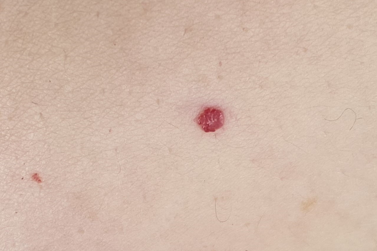
A ruby angioma, also called ruby hemangioma, cherry angioma, senile angioma or Campbell de Morgan angioma (the first British surgeon who studied and codified it in the first half of the 1800s), is a solid relief of the skin that is bright red in colour, hence the names ‘ruby’ and ‘cherry’.
This colouration is determined by the presence of capillary blood vessels that clump together inside.
Generally, the evolution of cherry angioma is very gradual
At the beginning, the size of the angioma is extremely small, about the size of a pinhead.
Subsequently, they increase in size, usually to about 3-5 millimetres or, more rarely, one centimetre.
They are not associated with any symptoms and do not give pain or any other local sensation such as itching or discomfort.
The patient does, however, have discomfort from the unsightliness associated with seeing their skin stained by red spots, especially when they are very numerous, and when they are localised in places such as the face.
They affect men and women indiscriminately after the age of 30 and often by familiarity
When they reach centimetres in size and are exophytic (detected on skin planes) or are located in rubbing sites, they can be easily traumatised, can become irritated and bleed profusely.
Conditions that may facilitate the occurrence of ruby angiomas are familiarity (reduced levels of microRNA427 have been found in the skin of patients with numerous of them, with associated overproduction of proteins that induce vascular proliferation), certain medications (cyclosporine and bromides), sunshine, age (increase in number after the age of 30; more than 75% of people over 75 would be carriers of at least one ruby angioma), hormonal pattern (increase in pregnancy), liver, pituitary and intestinal disorders.
They have no preference for a particular race, affecting men and women equally.
Although they can develop in any skin area of the human body, cherry angioma has a particular preference for the trunk, arms, legs and shoulders.
It has no negative consequences (it does not degenerate into a malignant tumour) and therefore does not require compulsory treatment for the patient’s health. However, if subject to repeated bleeding, or if it is perceived as an annoying blemish, the patient can safely ask the dermatologist for its removal.
Cherry angioma causes a red-purple spot on the skin
It is due to an abnormal development of the capillary cells in the dermis and epidermis.
The evolution of treatment possibilities compared to the past (surgery, cryotherapy, etc.) now makes it possible to remove them in a few minutes using laser technologies.
One of the lasers offering the best results is the NeodimioYAG, a laser consisting of a crystal of neodymium, yttrium, aluminium and garnet.
The energy of the vascular Nd-Yag (Neodymium YAG) laser, which emits a wavelength of 1064 nm, penetrates the tissue and selectively coagulates the blood without damaging the epidermis (principle of selective photo-thermolysis, which allows only the lesion to be coagulated).
Cooling the surface of the skin before, during and immediately after treatment allows the use of medium-high fluences that improve the coagulation results of the angioma, prevent damage to the epidermis and reduce discomfort for the patient.
A cherry angioma is a small, bright red relief on the skin
It may be single or multiple (several angiomas grouped adjacent to each other).
The ruby angioma removal session is quick, is performed on an outpatient basis, does not require anaesthesia and does not involve interruption of normal daily and work activities after the treatment.
The light flow of the Nd-Yag laser is directed directly onto the angioma to be removed
The patient experiences a slight, bearable and fleeting burning sensation. The ruby angioma undergoes a red to grey-black colour change, hardens and in the following days detaches without leaving a mark.
After the session the patient has no wound, no plaster and no local dressing.
Read Also
Emergency Live Even More…Live: Download The New Free App Of Your Newspaper For IOS And Android

Cavernous Angiomas: What They Are, How To Treat Them
Lymphoma: 10 Alarm Bells Not To Be Underestimated
Non-Hodgkin’s Lymphoma: Symptoms, Diagnosis And Treatment Of A Heterogeneous Group Of Tumours
CAR-T: An Innovative Therapy For Lymphomas
Acute Lymphoblastic Leukaemia: Long-Term Outcomes Described For Childhood ALL Survivors
Lymphangiomas And Lymphatic Malformations: What They Are, How To Treat Them
Melanoma: Prevention And Dermatological Examinations Are Essential Against Skin Cancer
Nail Melanoma: Prevention And Early Diagnosis
Dermatological Examination For Checking Moles: When To Do It
What Is A Tumour And How It Forms
Rare Diseases: New Hope For Erdheim-Chester Disease
How To Recognise And Treat Melanoma
Moles: Knowing Them To Recognise Melanoma
Skin Melanoma: Types, Symptoms, Diagnosis And The Latest Treatments
Nevi: What They Are And How To Recognise Melanocytic Moles
Bluish Color Of Baby’s Skin: Could Be Tricuspid Atresia
Skin Diseases: Xeroderma Pigmentosum
Basal Cell Carcinoma, How Can It Be Recognised?
Autoimmune Diseases: Care And Treatment Of Vitiligo
Epidermolysis Bullosa And Skin Cancers: Diagnosis And Treatment
SkinNeutrAll®: Checkmate For Skin-Damaging And Flammable Substances
Healing Wounds And Perfusion Oximeter, New Skin-Like Sensor Can Map Blood-Oxygen Levels
Psoriasis, An Ageless Skin Disease
Psoriasis: It Gets Worse In Winter, But It’s Not Just The Cold That’s To Blame
Childhood Psoriasis: What It Is, What The Symptoms Are And How To Treat It
Topical Treatments For Psoriasis: Recommended Over-The-Counter And Prescription Options
What Are The Different Types Of Psoriasis?
Phototherapy For The Treatment Of Psoriasis: What It Is And When It Is Needed
Skin Diseases: How To Treat Psoriasis?
Skin Cancers: Prevention And Care